. 2020 Aug 23;12(9):925.
doi: 10.3390/v12090925.

N-glycosylation in the Pre-Membrane Protein Is Essential for the Zika Virus Life Cycle

Affiliations

N-glycosylation in the Pre-Membrane Protein Is Essential for the Zika Virus Life Cycle

Yong-Dae Gwon et al. Viruses. .

Abstract

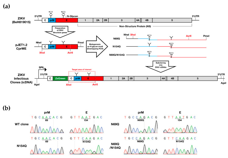
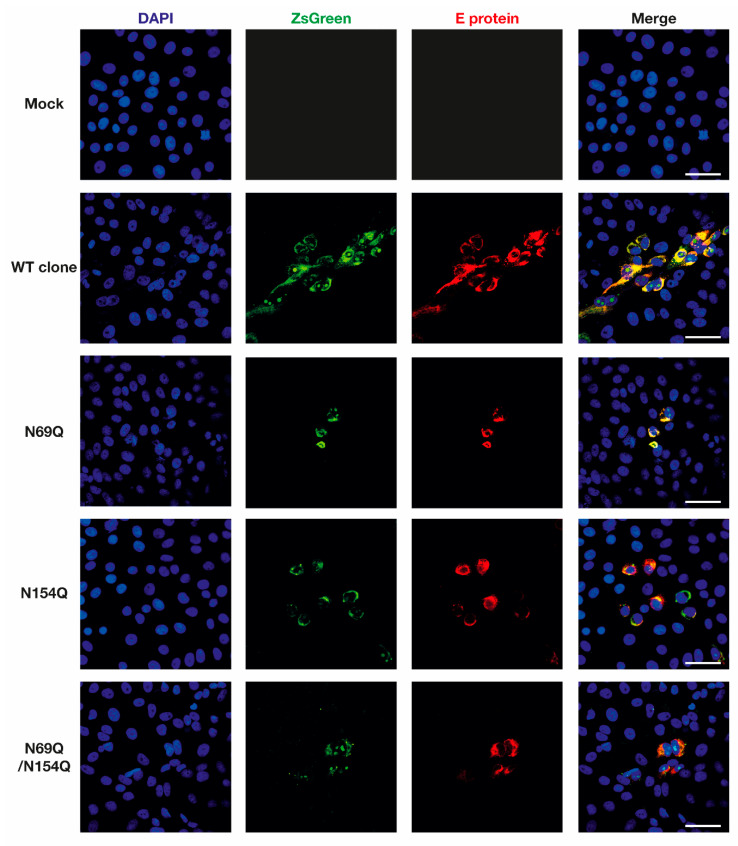
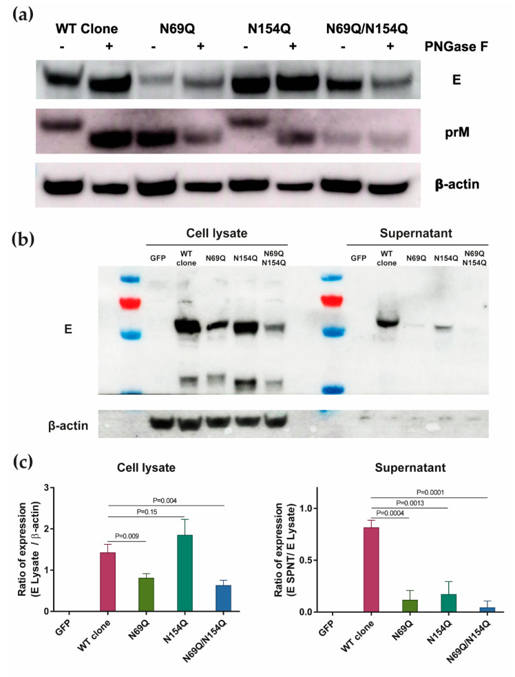
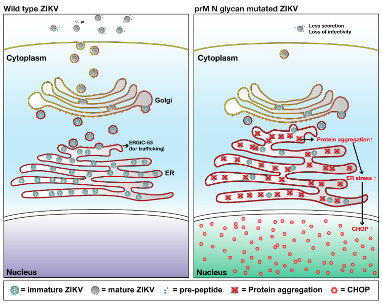
Asparagine (N)-linked protein glycosylation plays an important role in protein synthesis and modification. Two Zika virus (ZIKV) structural proteins, the pre-membrane (prM) and envelope (E) protein are N-glycosylated. The prM protein of all ZIKV strains contains a single N-linked glycosylation site, while not all strains contain an N-linked site in the E protein. Our aim was to examine the impact of prM and E N-linked glycosylation on ZIKV infectivity and cell trafficking. Using a ZIKV infectious clone, we found that when the N-glycan sites were removed, the prM- and the prM/E-double mutants did not produce an infectious virus in the supernatant. Further, by using ZIKV prME constructs, we found that N-glycosylation was necessary for effective secretion of ZIKV virions. The absence of the N-glycan on prM or E caused protein aggregation in the rough endoplasmatic reticulum (ER) compartment. The aggregation was more pronounced for the prM-mutation, and the mutant virus lost the ER-Golgi intermediate compartment (ERGIC) localization. In addition, lack of the N-glycan on prM induced nuclear translocation of CCAAT-enhancer-binding protein homologous protein (CHOP), an ER stress marker. To conclude, we show that the prM N-glycan is essential for the ZIKV infectious cycle, and plays an important role in viral protein trafficking, protein folding, and virion assembly.

Keywords: N-glycosylation; Zika virus; envelope; pre-membrane; virus life cycle.


Conflict of interest statement

The authors declare no conflict of interest.

Figures

Figure 1
Figure 1
Generating N-glycosylation mutants of ZIKV BeH819015 in prM and/or E. (a) The structural genes of ZIKV BeH819015 were cloned into a pJET1.2 vector and N-glycan motif mutagenesis was introduced by overlapping polymerase chain reaction (PCR). Mutated PCR amplicons were sub-cloned into the infectious clone of ZIKV BeH819015 that contained a ZsGreen marker by using the indicated restriction endonuclease sites (red color). SP6 promoter sequence is present upstream of sequence corresponding to ZIKV genome; (b) Sequence chromatograms of the prM and E protein glycosylation motifs of ZIKV wild-type (WT), N69Q, N154Q or N69Q/N154Q clones. ZIKV = Zika virus; UTR = untranslated region; C = capsid protein; prM = pre-membrane protein; E = envelope protein; ZsGreen = green fluorescent protein derived from zoanthus species; WT clone = wild-type ZIKV with the ZsGreen reporter gene; N69Q = ZIKV with a mutated N-glycosylation site in prM; N154Q = ZIKV with a mutated N-glycosylation site in E; N69Q/N154Q = ZIKV with mutated N-glycosylation sites in prM and E.
Figure 2
Figure 2
Confirmation of the inserted-marker (ZsGreen) and ZIKV E protein expression from rescued ZIKV variants. Vero B4 cells were grown on eight-well chamber slides. At 50% confluence, the cells were transfected with in vitro transcribed ZIKV RNA. At 72 hpt, the cells were fixed and stained with rabbit anti-ZIKV E antibody, anti-rabbit conjugated to Alexa Fluor 568 and DAPI. The samples were analyzed using a Nikon A1R+ confocal microscope and ImageJ software. Scale bar = 50 µm. DAPI = 4′,6-diamidino-2-phenylindole; ZsGreen = green fluorescent protein derived from Zoanthus sp.; E protein = envelope protein; WT clone = wild-type ZIKV with the ZsGreen reporter gene; N69Q = ZIKV with a mutated N-glycosylation site in prM; N154Q = ZIKV with a mutated N-glycosylation site in E; N69Q/N154Q = ZIKV with mutated N-glycosylation sites in prM and E.
Figure 3
Figure 3
The N-glycan on ZIKV prM is important for the virus life cycle. (a) Schematic diagram of experiments. (b) Representative picture of ZsGreen expression from four variants of ZIKV RNA transfected into Vero B4 cells. The monitoring was performed as scheduled in Figure 3a. Scale bar = 20 µm. (c) Virus egress kinetics by qRT-PCR, depicted as copy number of ZIKV NS5 RNA in 100 µL of harvested supernatant, as scheduled in Figure 3a. Mean values and standard deviations are shown from three independent experiments. (d) Representative picture of the plaque-forming ability of harvested viruses. The ZIKV strains MR766 and BeH819015 (approximately, 100 plaque-forming units) were used as positive controls. Respectively, virus titer was measured using plaque assay and the average of virus titer and standard deviations are shown from two independent experiments. (e) Confirmation of presence/absence of the N-glycan sites in the prM and E proteins. Vero B4 cells were infected with supernatant from day 6 (Figure 3a). Three days after infection, the cell lysate was harvested and used for prM and E protein detection by Western blotting, with either absence or presence of peptide N-glycosidase F (PNGase F) treatment. The ZIKV strains MR766 (absence of N-glycan in E) and BeH819015 (presence of N-glycan in E) were used as controls.
Figure 4
Figure 4
The lack of N-glycan on the pre-membrane (prM) or/and E protein caused impairment in the E protein expression and secretion. (a) Confirmation of absence/presence of N-glycosylation in the prM- and E proteins expressed from constructed plasmids. For the Western blotting, proteins were stained with anti-ZIKV E -, anti-ZIKV prM- or anti-β actin protein antibody (b) Representative Western blot picture of E- and β-actin protein expression in harvested cell lysate and cell culture supernatant from three independent experiments. (c) Quantification of E protein expression with Image J. To normalize the E protein expression in the cell lysate, the expression level of E protein was divided by the expression level of β-actin. To normalize the E protein expression in the supernatant, the expression level of E protein from the supernatant was divided by the expression level of E protein from the cell lysate. The experiment was repeated three times, average and SD are shown. Statistical significance was determined by two-tailed t-test. p = p value. GFP = pcDNA3.1-GFP vector; WT clone = pcDNA3.1-prME vectors with wild-type ZIKV prM-E genes; N69Q = pcDNA3.1-prME vector with a mutated N-glycosylation site in prM; N154Q = pcDNA3.1-prME vector with a mutated N-glycosylation site in E; N69Q/N154Q = pcDNA3.1-prME vector with mutated N-glycosylation sites in prM and E; PNGase F = Peptide:N-glycosidase F.
Figure 5
Figure 5
Co-localization of the E protein expression with various cellular protein markers. Vero cells were transfected with pcDNA3.1-prME plasmids. (a) The immunofluorescence microscopy images of ZIKV E-Sec61α double-stained Vero B4 cells. After 48 h, cells were stained with a rabbit anti-ZIKV E protein antibody and a mouse anti-Sec61 alpha antibody followed by secondary antibody and DAPI. (b) The immunofluorescence microscopy images of ZIKV E-ERGIC (ER-Golgi intermediate compartment) 53 double-stained Vero B4 cells. After 48 h, cells were stained with a rabbit anti-ZIKV E protein antibody and a mouse anti-ERGIC 53 antibody, followed by secondary antibody and DAPI. The samples were analyzed using a Zeiss 710 confocal microscope and ImageJ software. Scale bar = 20 µm. Sec61α = Protein transport protein Sec61 subunit alpha; ERGIC 53 = ER-Golgi intermediate compartment 53 kDa protein or lectin mannose-binding 1.
Figure 6
Figure 6
The lack of N-glycan on the prME protein induced ER stress. For the immunofluorescence assay, Vero cells were transfected with pcDNA3.1-prME plasmids for 48 h, samples were stained with a mouse anti-CHOP (CCAAT-enhancer-binding protein homologous protein) antibody together with a rabbit anti-ZIKV E antibody followed by secondary antibodies and DAPI. In positive controls, CHOP expression was induced with Brefeldin A (1 µg/mL) or Tunicamycin (1 µg/mL) for 24 h. Representative images of ZIKV E-CHOP double-stained Vero B4 cells. Scale bar = 20 µm.
Figure 7
Figure 7
Hypothetical model of the role of N-glycosylation of the prM protein in ZIKV. Mutation in the prM N-glycosylation site caused accumulation of virus proteins in ER, which induced increased ER stress in the cell. Eventually, the up-regulated ER stress triggered the expression of CHOP and its translocation into nucleus. ZIKV = Zika virus; Golgi = golgi apparatus; ER = endoplasmic reticulum; ERGIC-53 = ER-Golgi intermediate compartment 53 kDa protein or lectin mannose-binding 1; prM = pre-membrane protein; CHOP = C/EBP Homologous Protein.

References

    1. Gould E.A., Solomon T. Pathogenic flaviviruses. Lancet. 2008;371:500–509. doi: 10.1016/S0140-6736(08)60238-X. - DOI - PubMed
    1. Hamel R., Dejarnac O., Wichit S., Ekchariyawat P., Neyret A., Luplertlop N., Perera-Lecoin M., Surasombatpattana P., Talignani L., Thomas F., et al. Biology of Zika Virus Infection in Human Skin Cells. J. Virol. 2015;89:8880–8896. doi: 10.1128/JVI.00354-15. - DOI - PMC - PubMed
    1. Holbrook M.R. Historical Perspectives on Flavivirus Research. Viruses. 2017;9:97. doi: 10.3390/v9050097. - DOI - PMC - PubMed
    1. Taguwa S., Yeh M.T., Rainbolt T.K., Nayak A., Shao H., Gestwicki J.E., Andino R., Frydman J. Zika Virus Dependence on Host Hsp70 Provides a Protective Strategy against Infection and Disease. Cell Rep. 2019;26:906–920. doi: 10.1016/j.celrep.2018.12.095. - DOI - PMC - PubMed
    1. Musso D., Ko A.I., Baud D. Zika Virus Infection—After the Pandemic. N. Engl. J. Med. 2019;381:1444–1457. doi: 10.1056/NEJMra1808246. - DOI - PubMed

Publication types

Substances