

A Surrogate Animal Model for Screening of Ebola and Marburg Glycoprotein-Targeting Drugs Using Pseudotyped Vesicular Stomatitis Viruses
- PMID: 32842671
- PMCID: PMC7552044
- DOI: 10.3390/v12090923
A Surrogate Animal Model for Screening of Ebola and Marburg Glycoprotein-Targeting Drugs Using Pseudotyped Vesicular Stomatitis Viruses
Abstract
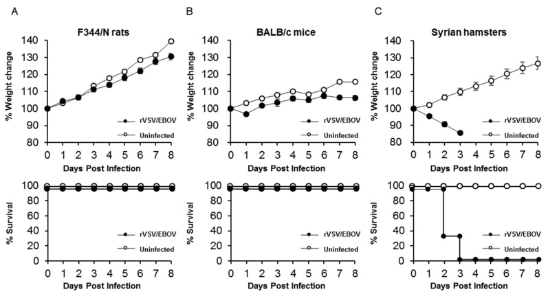
Filoviruses, including Ebola virus (EBOV) and Marburg virus (MARV), cause severe hemorrhagic fever in humans and nonhuman primates with high mortality rates. There is no approved therapy against these deadly viruses. Antiviral drug development has been hampered by the requirement of a biosafety level (BSL)-4 facility to handle infectious EBOV and MARV because of their high pathogenicity to humans. In this study, we aimed to establish a surrogate animal model that can be used for anti-EBOV and -MARV drug screening under BSL-2 conditions by focusing on the replication-competent recombinant vesicular stomatitis virus (rVSV) pseudotyped with the envelope glycoprotein (GP) of EBOV (rVSV/EBOV) and MARV (rVSV/MARV), which has been investigated as vaccine candidates and thus widely used in BSL-2 laboratories. We first inoculated mice, rats, and hamsters intraperitoneally with rVSV/EBOV and found that only hamsters showed disease signs and succumbed within 4 days post-infection. Infection with rVSV/MARV also caused lethal infection in hamsters. Both rVSV/EBOV and rVSV/MARV were detected at high titers in multiple organs including the liver, spleen, kidney, and lungs of infected hamsters, indicating acute and systemic infection resulting in fatal outcomes. Therapeutic effects of passive immunization with an anti-EBOV neutralizing antibody were specifically observed in rVSV/EBOV-infected hamsters. Thus, this animal model is expected to be a useful tool to facilitate in vivo screening of anti-filovirus drugs targeting the GP molecule.
Keywords: Ebola virus; Filovirus; Marburg virus; Syrian hamster; animal model; drug screening; recombinant vesicular stomatitis virus.
Conflict of interest statement
The authors declare that the research was conducted in the absence of any commercial or financial relationships that could be construed as a potential conflict of interest.
Figures
Similar articles
-
Single-Dose Trivalent VesiculoVax Vaccine Protects Macaques from Lethal Ebolavirus and Marburgvirus Challenge.J Virol. 2018 Jan 17;92(3):e01190-17. doi: 10.1128/JVI.01190-17. Print 2018 Feb 1. J Virol. 2018. PMID: 29142131 Free PMC article.
-
Establishment and application of a surrogate model for human Ebola virus disease in BSL-2 laboratory.Virol Sin. 2024 Jun;39(3):434-446. doi: 10.1016/j.virs.2024.03.010. Epub 2024 Mar 29. Virol Sin. 2024. PMID: 38556051 Free PMC article.
-
Recombinant Protein Filovirus Vaccines Protect Cynomolgus Macaques From Ebola, Sudan, and Marburg Viruses.Front Immunol. 2021 Aug 18;12:703986. doi: 10.3389/fimmu.2021.703986. eCollection 2021. Front Immunol. 2021. PMID: 34484200 Free PMC article.
-
Recombinant vesicular stomatitis virus-based vaccines against Ebola and Marburg virus infections.J Infect Dis. 2011 Nov;204 Suppl 3(Suppl 3):S1075-81. doi: 10.1093/infdis/jir349. J Infect Dis. 2011. PMID: 21987744 Free PMC article. Review.
-
Ebola and Marburg virus vaccines.Virus Genes. 2017 Aug;53(4):501-515. doi: 10.1007/s11262-017-1455-x. Epub 2017 Apr 26. Virus Genes. 2017. PMID: 28447193 Free PMC article. Review.
Cited by
-
Characterization of a Vesicular Stomatitis Virus-Vectored Recombinant Virus Bearing Spike Protein of SARS-CoV-2 Delta Variant.Microorganisms. 2023 Feb 8;11(2):431. doi: 10.3390/microorganisms11020431. Microorganisms. 2023. PMID: 36838396 Free PMC article.
-
Mining of Marburg Virus Proteome for Designing an Epitope-Based Vaccine.Front Immunol. 2022 Jul 15;13:907481. doi: 10.3389/fimmu.2022.907481. eCollection 2022. Front Immunol. 2022. PMID: 35911751 Free PMC article.
-
Pseudotyped Vesicular Stomatitis Virus-Severe Acute Respiratory Syndrome-Coronavirus-2 Spike for the Study of Variants, Vaccines, and Therapeutics Against Coronavirus Disease 2019.Front Microbiol. 2022 Jan 14;12:817200. doi: 10.3389/fmicb.2021.817200. eCollection 2021. Front Microbiol. 2022. PMID: 35095820 Free PMC article. Review.
-
Emerging and reemerging infectious diseases: global trends and new strategies for their prevention and control.Signal Transduct Target Ther. 2024 Sep 11;9(1):223. doi: 10.1038/s41392-024-01917-x. Signal Transduct Target Ther. 2024. PMID: 39256346 Free PMC article. Review.
-
A rabies virus-vectored vaccine expressing two copies of the Marburg virus glycoprotein gene induced neutralizing antibodies against Marburg virus in humanized mice.Emerg Microbes Infect. 2023 Dec;12(1):2149351. doi: 10.1080/22221751.2022.2149351. Emerg Microbes Infect. 2023. PMID: 36453198 Free PMC article.
References
Publication types
MeSH terms
Substances
LinkOut - more resources
Full Text Sources
Medical
