

Primary pulmonary cystic Echinococcus in an immunocompetent patient
- PMID: 32843399
- PMCID: PMC7449490
- DOI: 10.1136/bcr-2020-234578
Primary pulmonary cystic Echinococcus in an immunocompetent patient
Abstract
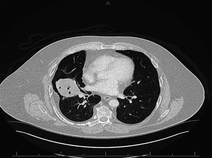
A 60-year-old man was referred to the interventional pulmonology clinic with a large right-sided intraparenchymal lung mass and a second, smaller lesion in the left lower lobe, accompanied by intermittent haemoptysis, fever, chills, productive cough of white phlegm as well as dizziness and weakness. He had presented previously and was being evaluated for the possibility of malignancy. Investigations had revealed 'hooklets' (protoscolices) of hydatid cysts, most likely representing the parasite Echinococcus Successful surgical excision of the affected lobe, lung decortication, partial pleurectomy and pneumolysis of the adhesions was performed, along with long-term antiparasitic therapy. The initial differential diagnosis for this patient was challenging and required multimodal investigations. The patient made good recovery and continued to be followed by infectious disease specialists for management of antiparasitic therapy.
Keywords: infectious diseases; medical management; respiratory medicine.
© BMJ Publishing Group Limited 2020. No commercial re-use. See rights and permissions. Published by BMJ.
Conflict of interest statement
Competing interests: None declared.
Figures


References
-
- Center for Disease Control and Prevention Parasites – echinococcosis, 2012.
-
- Gru B, Schmidgerger J, Drews O, et al. Imaging in alveolar echinococcosis (AE): comparison of Echinococcus multilocularis classification for computed-tomography (EMUC-CT) and ultrasonography (EMUC-US). Radiol Infect Dis 2017;4:70–7.
Publication types
MeSH terms
Substances
LinkOut - more resources
Full Text Sources