

Rotavirus Infection and Cytopathogenesis in Human Biliary Organoids Potentially Recapitulate Biliary Atresia Development
- PMID: 32843549
- PMCID: PMC7448284
- DOI: 10.1128/mBio.01968-20
Rotavirus Infection and Cytopathogenesis in Human Biliary Organoids Potentially Recapitulate Biliary Atresia Development
Abstract
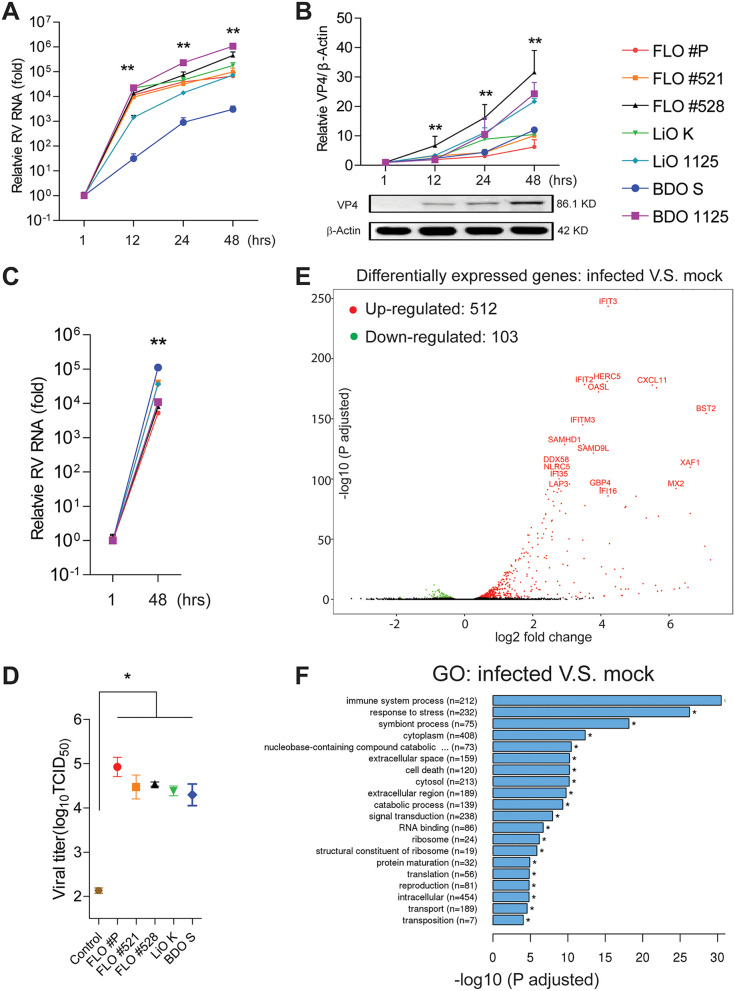
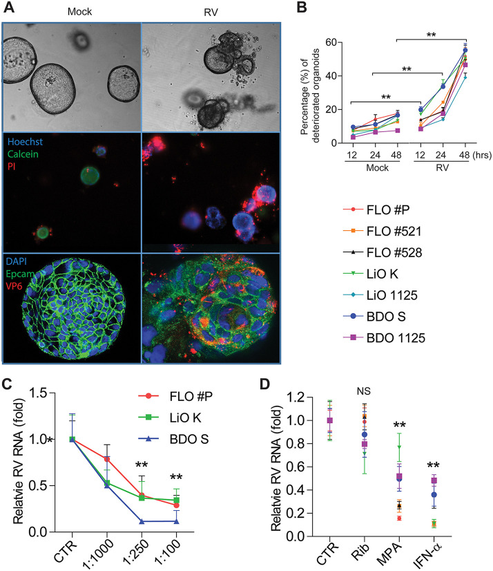
Biliary atresia (BA) is a neonatal liver disease characterized by progressive fibroinflammatory obliteration of both intrahepatic and extrahepatic bile ducts. The etiologies of BA remain largely unknown, but rotavirus infection has been implicated at least for a subset of patients, and this causal relation has been well demonstrated in mouse models. In this study, we aim to further consolidate this evidence in human biliary organoids. We obtained seven batches of human biliary organoids cultured from fetal liver, adult liver, and bile duct tissues. We found that these organoids are highly susceptible and support the full life cycle of rotavirus infection in three-dimensional culture. The robust infection triggers active virus-host interactions, including interferon-based host defense mechanisms and injury responses. We have observed direct cytopathogenesis in organoids upon rotavirus infection, which may partially recapitulate the development of BA. Importantly, we have demonstrated the efficacy of mycophenolic acid and interferon alpha but not ribavirin in inhibiting rotavirus in biliary organoids. Furthermore, neutralizing antibody targeting rotavirus VP7 protein effectively inhibits infection in organoids. Thus, we have substantiated the causal evidence of rotavirus inducing BA in humans and provided potential strategies to combat the disease.IMPORTANCE There is substantial evidence indicating the possible involvement of rotavirus in biliary atresia (BA) development, at least in a subset of patients, but concrete proof remains lacking. In a mouse model, it has been well demonstrated that rotavirus can infect the biliary epithelium to cause biliary inflammation and obstruction, representing the pathogenesis of BA in humans. By using recently developed organoids technology, we now have demonstrated that human biliary organoids are susceptible to rotavirus infection, and this provokes active virus-host interactions and causes severe cytopathogenesis. Thus, our model recapitulates some essential aspects of BA development. Furthermore, we have demonstrated that antiviral drugs and neutralizing antibodies are capable of counteracting the infection and BA-like morphological changes, suggesting their potential for mitigating BA in patients.
Keywords: biliary atresia; human organoids; rotavirus infection.
Copyright © 2020 Chen et al.
Figures
References
-
- Riepenhoff-Talty M, Gouvea V, Evans MJ, Svensson L, Hoffenberg E, Sokol RJ, Uhnoo I, Greenberg SJ, Schakel K, Zhaori G, Fitzgerald J, Chong S, El-Yousef M, Nemeth A, Brown M, Piccoli D, Hyams J, Ruffin D, Rossi T. 1996. Detection of group C rotavirus in infants with extrahepatic biliary atresia. J Infect Dis 174:8–15. doi: 10.1093/infdis/174.1.8. - DOI - PubMed
-
- Yin Y, Bijvelds M, Dang W, Xu L, van der Eijk AA, Knipping K, Tuysuz N, Dekkers JF, Wang Y, de Jonge J, Sprengers D, van der Laan LJW, Beekman JM, Ten Berge D, Metselaar HJ, de Jonge H, Koopmans MPG, Peppelenbosch MP, Pan Q. 2015. Modeling rotavirus infection and antiviral therapy using primary intestinal organoids. Antiviral Res 123:120–131. doi: 10.1016/j.antiviral.2015.09.010. - DOI - PubMed
Publication types
MeSH terms
Substances
LinkOut - more resources
Full Text Sources
Medical
