. 2020 Nov;69(11):2446-2457.
doi: 10.2337/db20-0579. Epub 2020 Aug 25.

YAP Activation in Renal Proximal Tubule Cells Drives Diabetic Renal Interstitial Fibrogenesis

Affiliations

YAP Activation in Renal Proximal Tubule Cells Drives Diabetic Renal Interstitial Fibrogenesis

Jianchun Chen et al. Diabetes. 2020 Nov.

Abstract

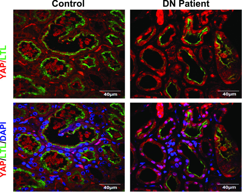
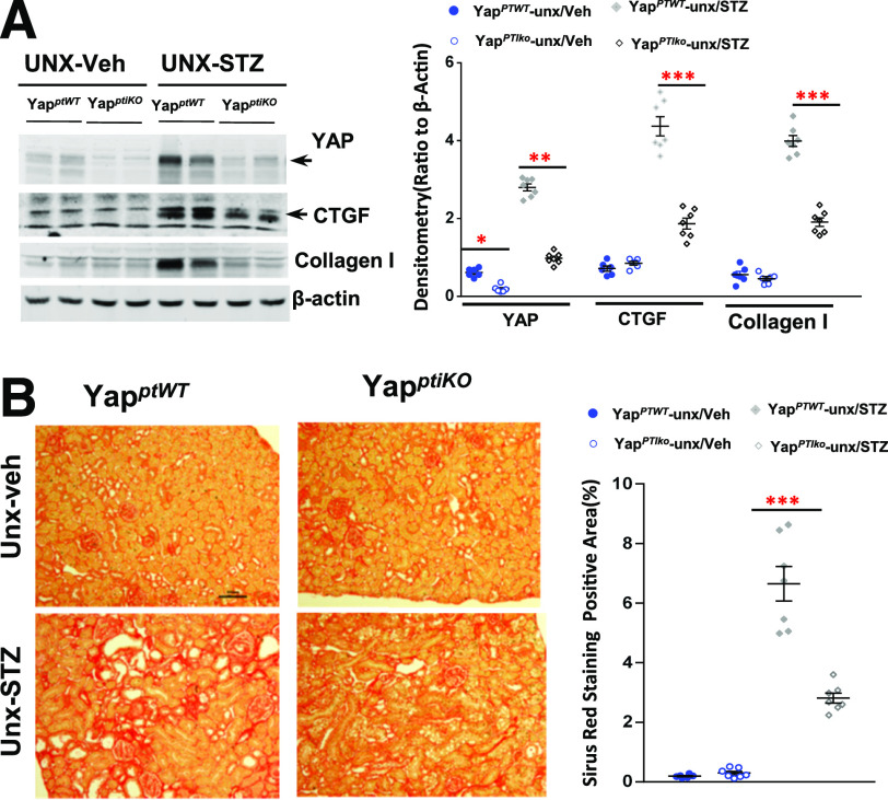
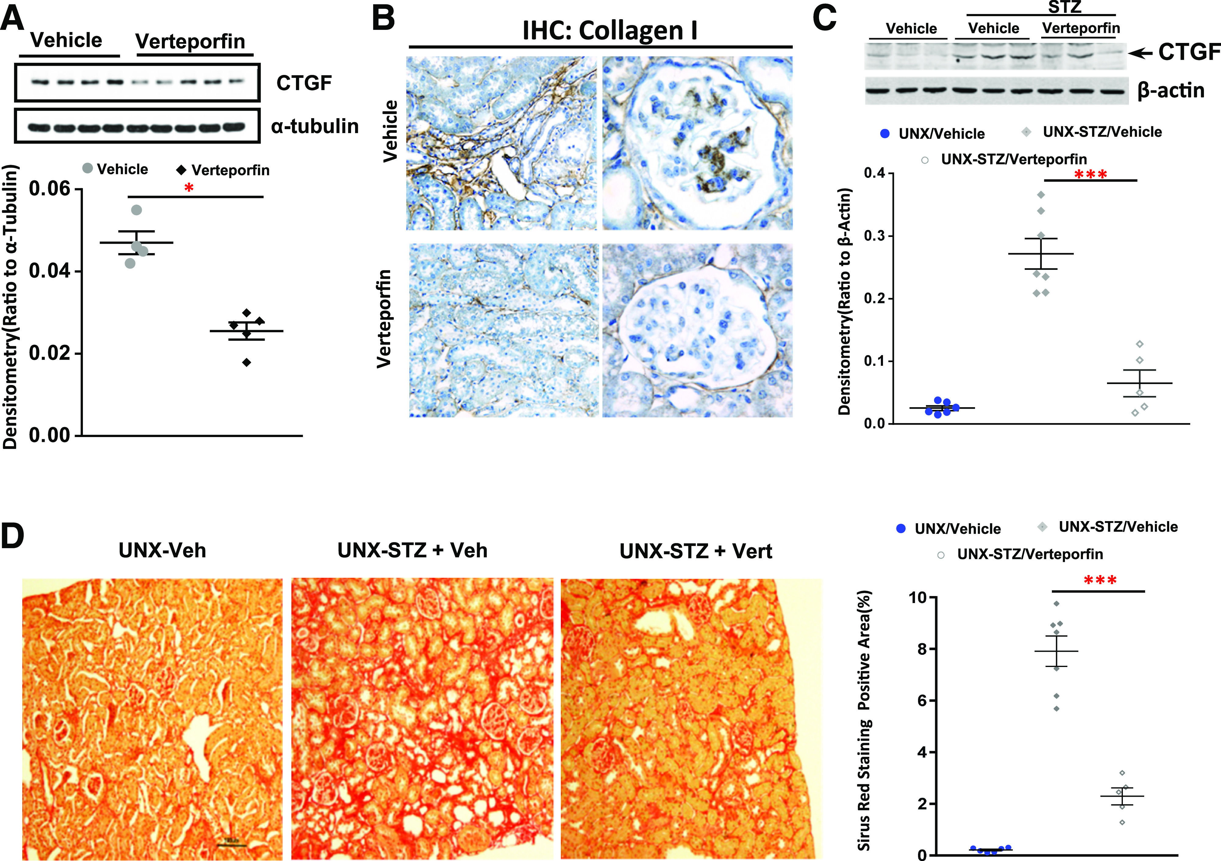
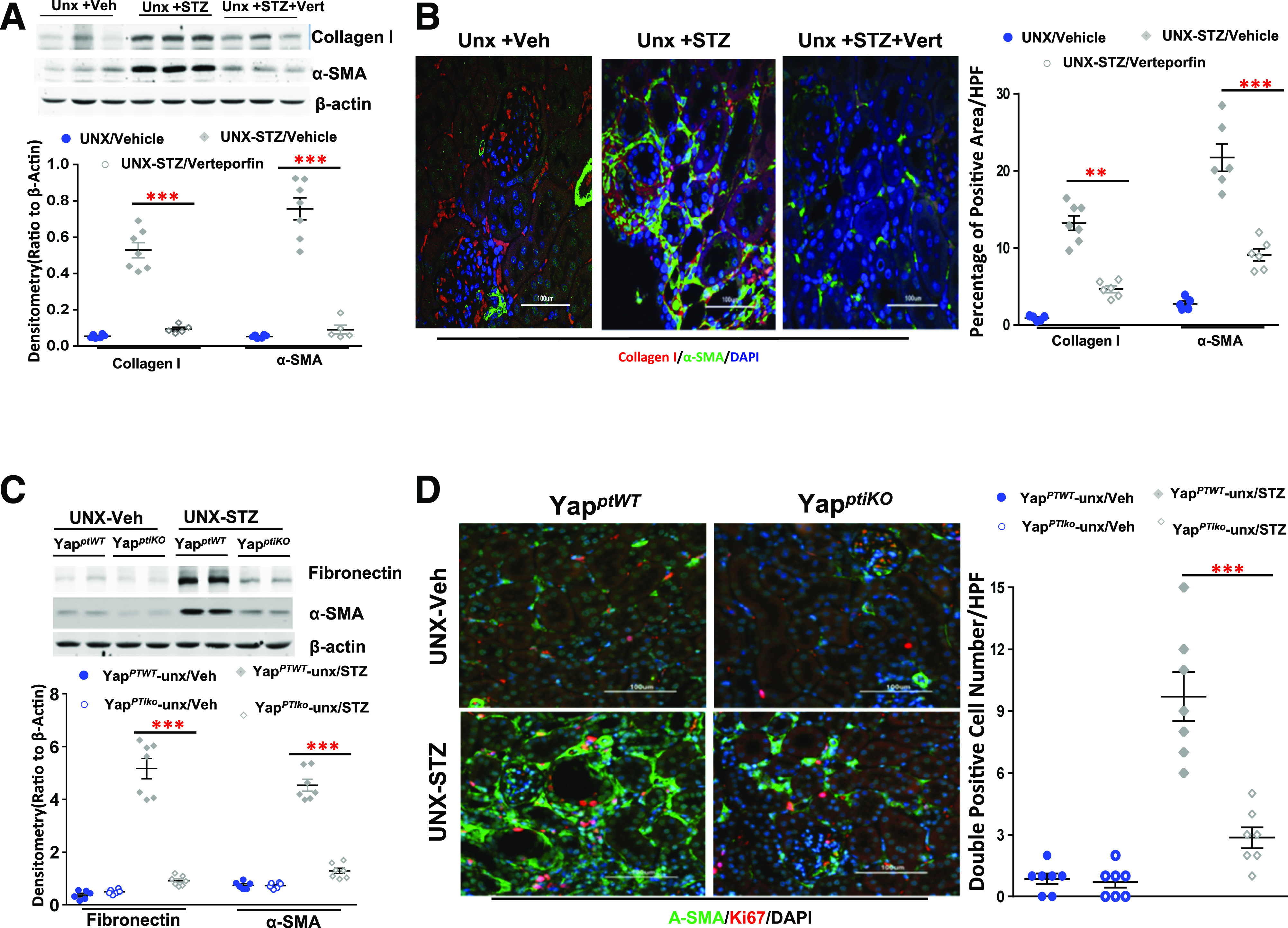
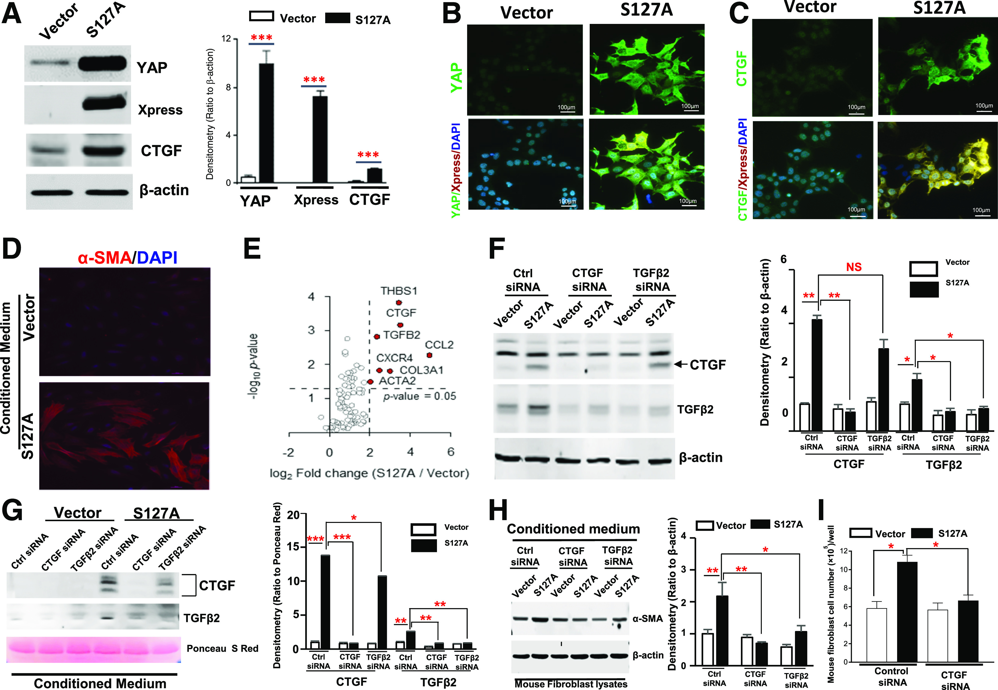
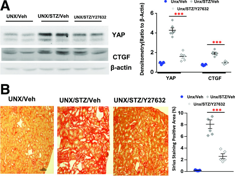
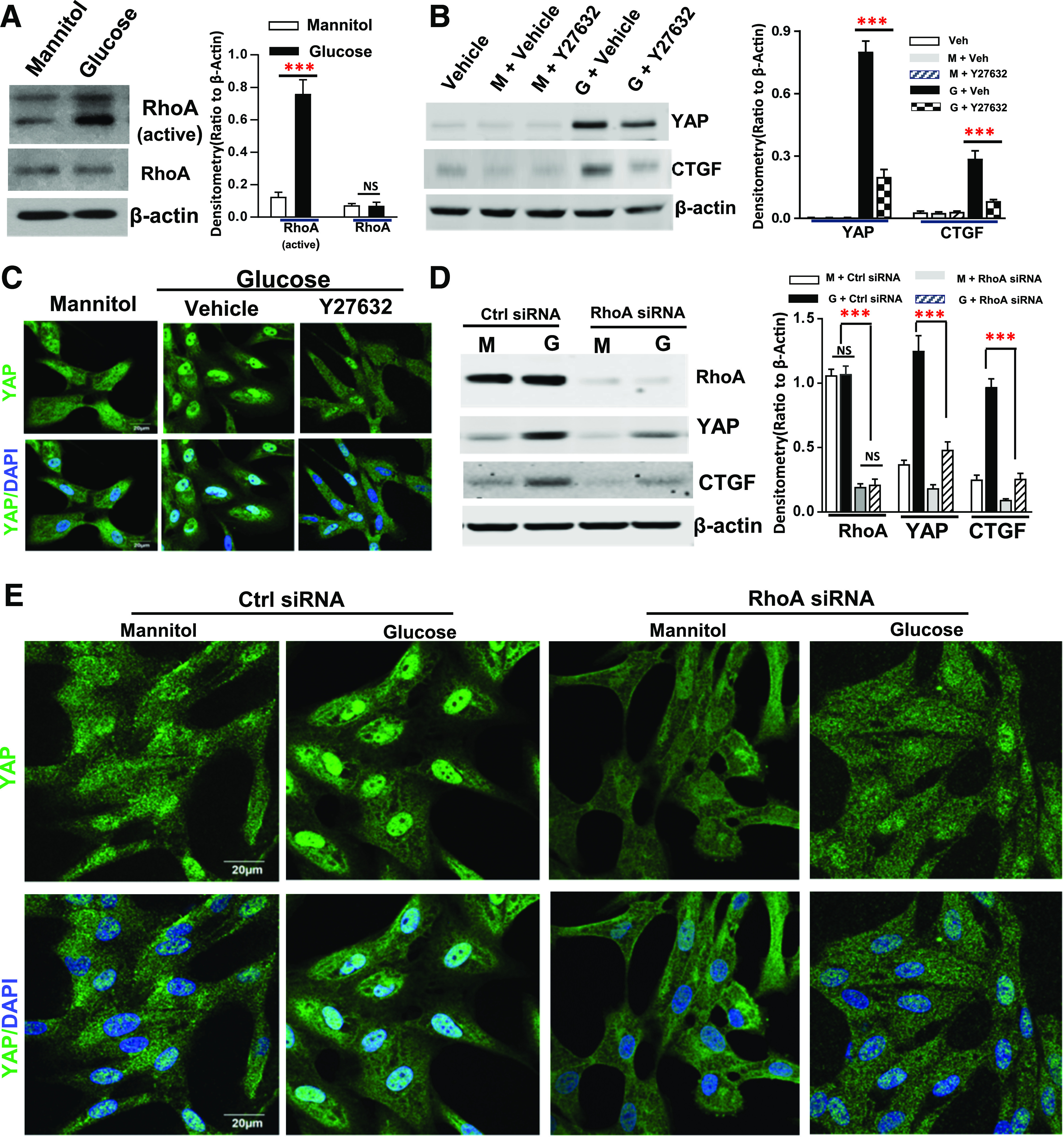
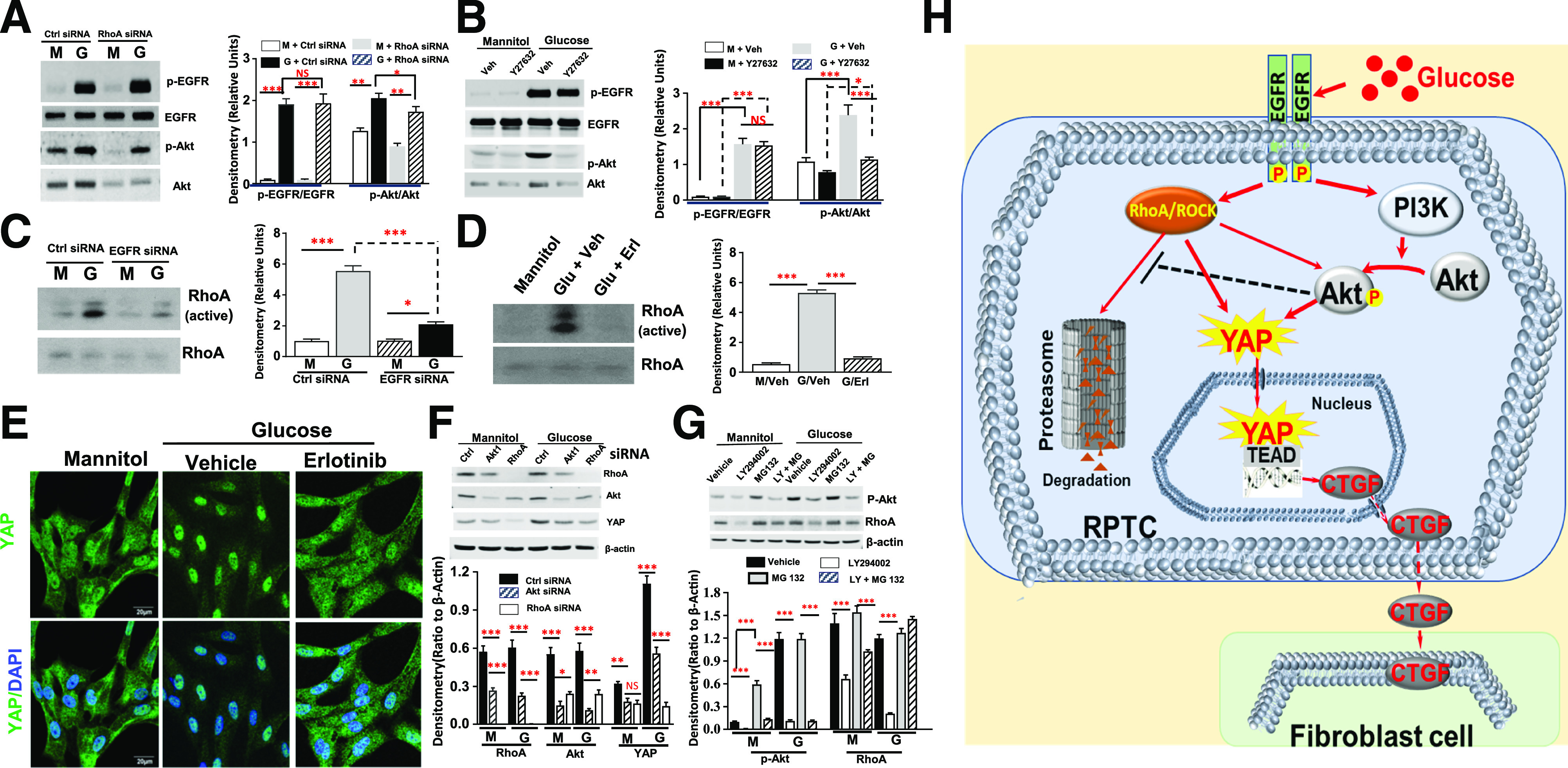
An increasing number of studies suggest that the renal proximal tubule is a site of injury in diabetic nephropathy (DN), and progressive renal tubulointerstitial fibrosis is an important mediator of progressive kidney dysfunction in DN. In this study, we observed increased expression and activation of YAP (yes-associated protein) in renal proximal tubule epithelial cells (RPTC) in patients with diabetes and in mouse kidneys. Inducible deletion of Yap specifically in RPTC or administration of the YAP inhibitor verteporfin significantly attenuated diabetic tubulointerstitial fibrosis. EGFR-dependent activation of RhoA/Rock and PI3K-Akt signals and their reciprocal interaction were upstream of proximal tubule YAP activation in diabetic kidneys. Production and release of CTGF in culture medium were significantly augmented in human embryonic kidney (HEK)-293 cells transfected with a constitutively active YAP mutant, and the conditioned medium collected from these cells activated and transduced fibroblasts into myofibroblasts. This study demonstrates that proximal tubule YAP-dependent paracrine mechanisms play an important role in diabetic interstitial fibrogenesis; therefore, targeting Hippo signaling may be a therapeutic strategy to prevent the development and progression of diabetic interstitial fibrogenesis.


Figures

Figure 1
Figure 1
YAP expression and nuclear distribution were increased in RPTC of kidneys in patients with diabetes. Representative IF images of control biopsy kidney specimens from patients without diabetes and patients with diabetes (n = 5) (red, YAP; green, LTL, an RPTC marker; blue, DAPI) (original magnification ×600).
Figure 2
Figure 2
YAP deletion specific in RPTC ameliorated interstitial fibrosis in diabetic mouse kidneys. A: Upregulation of expression of YAP, CTGF, and collagen I in the UNX STZ type 1 diabetic mouse cortical tissue was attenuated in YapPTiKO mice. B: Representative images of Sirius Red staining indicated that Yap deletion specific in RPTC reduced diabetic interstitial fibrosis (original magnification ×200). Values are mean ± SEM (n = 4–7 for each group). *P < 0.05, **P < 0.01, ***P < 0.001.
Figure 3
Figure 3
Blocking YAP-TEAD association reduced interstitial fibrosis in diabetic mouse kidneys. Nine- to 10-week old STZ diabetic eNOS−/− mice (A and B) or unilateral nephrectomized diabetic FVB/NJ mice (C and D) were administered vehicle or verteporfin (Vert) (100 mg/kg i.p. every other day) for 20 weeks: immunoblotting analysis of CTGF expression in renal cortical tissue lysates (A and C), representative immunohistochemical (IHC) staining of collagen I expression in both interstitial area and glomeruli (original magnification ×400) (B), and representative histology data of Sirius Red staining (original magnification ×200) (D). Values are mean ± SEM (n = 5–10 for each group). Veh, vehicle. *P < 0.05, ***P < 0.001.
Figure 4
Figure 4
Verteporfin (Vert) or RPTC-specific YAP deletion reduced ECM and α-SMA expression in diabetic kidneys. A: Verteporfin inhibited upregulation of collagen I and α-SMA in unilateral nephrectomized diabetic FVB/NJ mouse renal cortical tissue lysates. B: Representative IF of verteporfin inhibition of collagen I and α-SMA (original magnification ×200). C: Fibronectin and α-SMA upregulation in diabetic kidneys were significantly inhibited in YapPTiKO mice. D: Representative IF indicated inhibition of α-SMA expression and Ki67-positive cells in the YapPTiKO mouse kidneys (original magnification ×200). Values are mean ± SEM (n = 4–10 for each group). **P < 0.01, ***P < 0.001. HPF, high power field.
Figure 5
Figure 5
Conditioned medium from hYAP1S127A-HEK–activated fibroblasts. Generation of a stable transfectant (S127A) of a constitutively active human YAP1 mutant gene in HEK-293 cells was confirmed by immunoblotting analysis (A) and IF staining (B and C) (original magnification ×400). D: Conditioned medium from hYAP1S127A-HEK cells altered mouse fibroblast cell morphology and upregulated α-SMA expression (original magnification ×400). E: Screening results of possible profibrotic factors released from the active YAP-transfected HEK cells by using RT2 Profiler human fibrosis PCR array. F: Upregulation of CTGF and TGFβ2 expression in hYAP1S127A-HEK was effectively inhibited by transfection of specific siRNA respectively, and transfection of CTGF siRNA also blocked expression of TGFβ2. G: CTGF and TGFβ2 were detected in the cultured medium from hYAP1S127A-HEK cells; expressions were effectively abolished by transfection of their specific siRNAs. Silencing CTGF and TGFβ2 expression in hYAP1S127A-HEK cells by siRNAs inhibited conditioned medium–induced α-SMA expression in the mouse fibroblasts (H) and fibroblast cell proliferation (I). S127A, hYAP1S127A-HEK; Vector, vector-transfected HEK-293 cells. Values are mean ± SEM (the data were from at least three separate experiments). Xpress: N-terminal tag of the expression vector. *P < 0.05, **P < 0.01, ***P < 0.001. NS, nonsignificant difference. Ctrl, control; S Red, Sirius Red.
Figure 6
Figure 6
Inhibiting RhoA/ROCK signaling prevented YAP activation and ameliorated interstitial fibrosis in diabetic mice. Administration of a RhoA/ROCK inhibitor, Y27632, to diabetic FVB/NJ mice inhibited YAP and CTGF expression in renal cortical tissue lysates (A) and significantly attenuated interstitial fibrosis (original magnification ×200) (B). Values are mean ± SEM (n = 4–7 for each group). ***P < 0.001. Veh, vehicle.
Figure 7
Figure 7
RhoA activation mediated YAP activation in response to high glucose treatment in cultured hRPTC. High glucose treatment increased RhoA activity but not its expression (A), and Y27632 inhibited YAP expression and nuclear translocation and CTGF expression in response to high glucose (original magnification ×600) (B and C). Transfection of RhoA siRNA attenuated upregulation of YAP and CTGF in response to high glucose treatment (D) and inhibited YAP nuclear translocation (E) in response to high glucose treatment (original magnification ×600). Values are mean ± SEM (the data were from at least three separate experiments). ***P < 0.001. Ctrl, control; G, glucose; M, mannitol; NS, nonsignificant difference; Veh, vehicle.
Figure 8
Figure 8
Interactions between PI3K-Akt and RhoA/ROCK mediated EGFR-dependent YAP activation in RPTC in response to high glucose. A: Inhibition of RhoA expression did not affect EGFR activation but significantly inhibited Akt expression and phosphorylation in response to high glucose treatment in hRPTC. B: Y27632 did not affect EGFR activation but also decreased Akt expression and phosphorylation in response to high glucose treatment in hRPTC. C: siRNA knockdown of EGFR did not reduce RhoA expression but inhibited its activation. D: The EGFR tyrosine kinase inhibitor erlotinib did not alter RhoA expression but inhibited its activation. E: Erlotinib inhibited YAP nuclear translocation in response to high glucose treatment in hRPTC (original magnification ×600). F: siRNA knockdown of Akt1 reduced RhoA protein expression, and siRNA knockdown expression of either Akt1 or RhoA inhibited YAP expression in response to high glucose in hRPTC. G: The PI3K inhibitor LY294002 treatment attenuated RhoA protein expression, which was reversed by treatment of the cells with a cell-permeable proteasome inhibitor, MG-132. H: In response to hyperglycemia, EGF receptor (EGFR) mediates RhoA/ROCK– and PI3K-Akt–dependent YAP activation and subsequent downstream target gene CTGF expression, which was released into peritubular space to activate fibroblast proliferation and transition to myofibroblasts. In addition, RhoA/ROCK activation is essential for induction of Akt expression and phosphorylation, and the resulting Akt activation inhibits RhoA protein degradation by proteasome system. *P < 0.05, **P < 0.01, ***P < 0.001. Ctrl, control; G, glucose; M, mannitol; Veh, vehicle.

References

    1. Martínez-Castelao A, Navarro-González JF, Górriz JL, de Alvaro F. The concept and the epidemiology of diabetic nephropathy have changed in recent years. J Clin Med 2015;4:1207–1216 - PMC - PubMed
    1. Atkins RC, Zimmet P. Diabetic kidney disease: act now or pay later. Kidney Int 2010;77:375–377 - PubMed
    1. Bonventre JV Can we target tubular damage to prevent renal function decline in diabetes? Semin Nephrol 2012;32:452–462 - PMC - PubMed
    1. Chevalier RL The proximal tubule is the primary target of injury and progression of kidney disease: role of the glomerulotubular junction. Am J Physiol Renal Physiol 2016;311:F145–F161 - PMC - PubMed
    1. Miltényi M, Körner A, Tulassay T, Szabó A. Tubular dysfunction in type I diabetes mellitus. Arch Dis Child 1985;60:929–931 - PMC - PubMed

Publication types

MeSH terms