. 2020 Aug 25;10(1):14151.
doi: 10.1038/s41598-020-70822-4.

TRPV4 activates the Cdc42/N-wasp pathway to promote glioblastoma invasion by altering cellular protrusions

Affiliations

TRPV4 activates the Cdc42/N-wasp pathway to promote glioblastoma invasion by altering cellular protrusions

Wei Yang et al. Sci Rep. .

Abstract

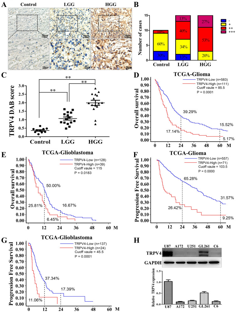
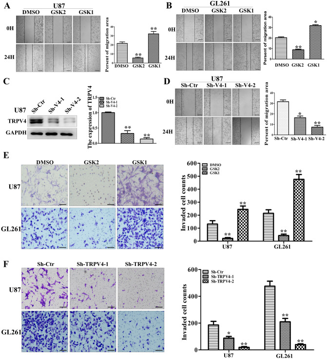
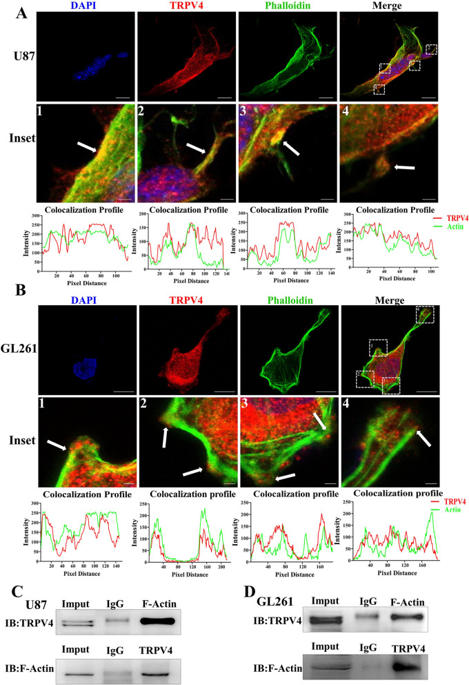
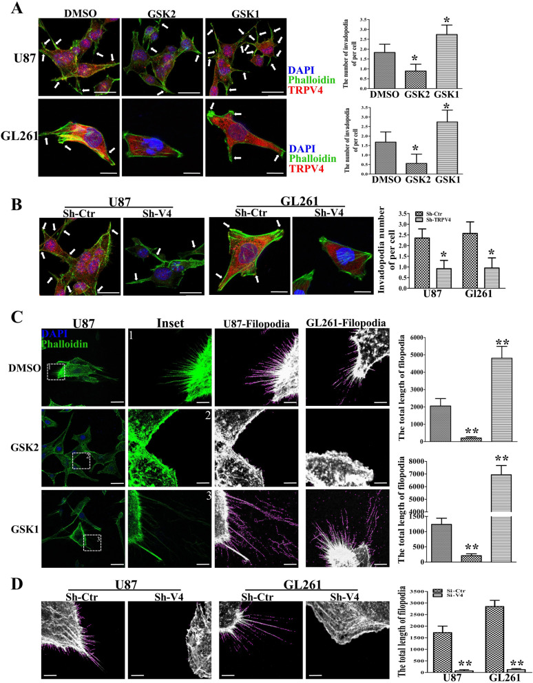
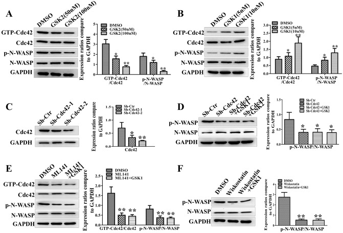
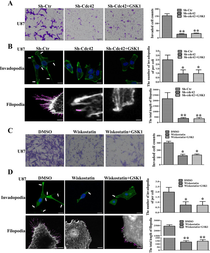
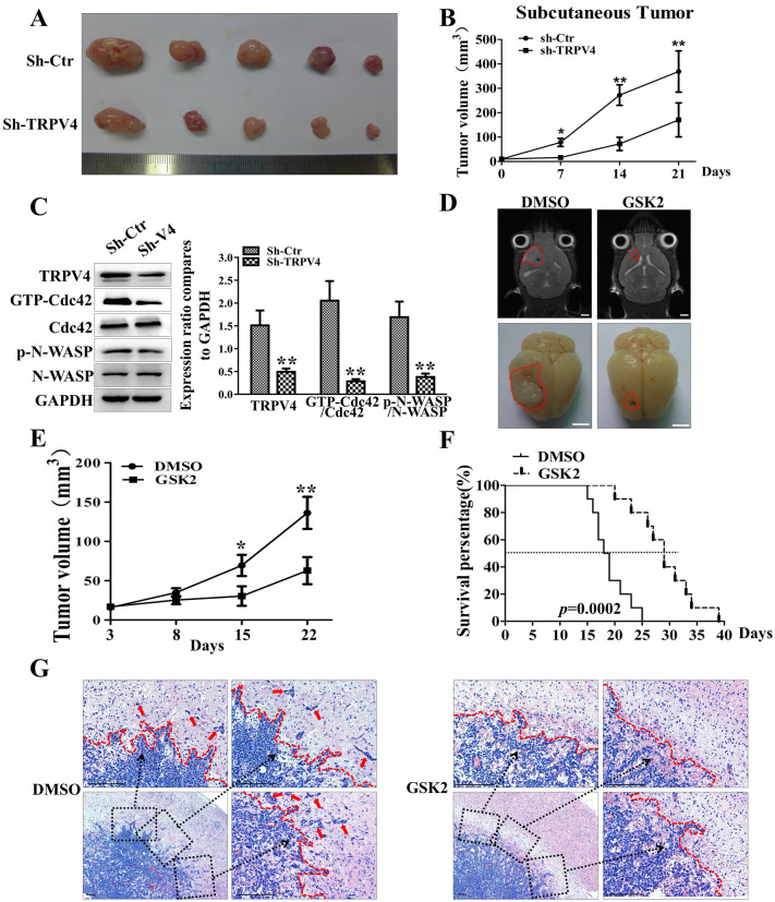
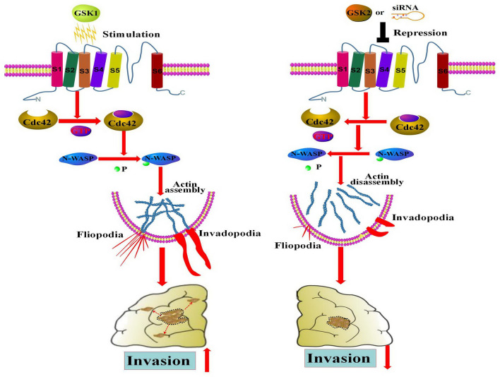
The invasion ability of glioblastoma (GBM) causes tumor cells to infiltrate the surrounding brain parenchyma and leads to poor outcomes. Transient receptor potential vanilloid 4 (TRPV4) exhibits a remarkable role in cancer cell motility, but the contribution of TRPV4 to glioblastoma metastasis is not fully understood. Here, we reported that TRPV4 expression was significantly elevated in malignant glioma compared to normal brain and low-grade glioma, and TRPV4 expression was negatively correlated with the prognosis of glioma patients. Functionally, stimulation of TRPV4 promoted glioblastoma cell migration and invasion, and repression of TRPV4 hindered the migration and invasion of glioblastoma cells in vitro. Molecularly, TRPV4 strongly colocalized and interacted with skeletal protein-F-actin at cellular protrusions, and TRPV4 regulated the formation of invadopodia and filopodia in glioblastoma cells. Furthermore, the Cdc42/N-wasp axis mediated the effect of TRPV4-regulated cellular protrusions and invasion. Foremost, TRPV4 inhibitor treatment or downregulation of TRPV4 significantly reduced the invasion-growth of subcutaneously and intracranially transplanted glioblastoma in mice. In conclusion, the TRPV4/Cdc42/wasp signaling axis regulates cellular protrusion formation in glioblastoma cells and influences the invasion-growth phenotype of glioblastoma in vivo. TRPV4 may serve as a prognostic factor and specific therapeutic target for GBM patients.


Conflict of interest statement

The authors declare no competing interests.

Figures

Figure 1
Figure 1
TRPV4 is highly expressed in malignant glioma, and higher expression indicates poor prognosis in glioma patients. (A) The expression of TRPV4 in normal brain tissues (control), low-grade glioma (LGG) and high-grade glioma (HGG) tissues was detected by IHC, scale bar = 50 µm or 20 µm. (B) The percentage of TRPV4 staining grades (− , + , +  + , +  + +) in controls, LGG and HGG. (C) IHC staining for TRPV4 was assessed semiquantitatively. TRPV4 intensity and the percentage of positive cells were multiplied to give a weighted score for each case. Data are plotted as the means ± SD from control (0.37 ± 0.14, n = 10), LGG (1.07 ± 0.36, n = 15) and HGG (1.99 ± 0.59, n = 15), **p < 0.01. (D) Kaplan–Meier analysis of the 2-year and 5-year overall survival (OS) rates of TRPV4low patients and TRPV4high patients from the TCGA-glioma dataset. (E) Kaplan–Meier analysis of the 1-year and 2-year OS rates of TRPV4low and TRPV4high patients from the TCGA-glioblastoma dataset. (F) Kaplan–Meier analysis of 2-year and 5-year progression free survival (PFS) rates of TRPV4low patients and TRPV4high patients from the TCGA-glioma dataset (G) Kaplan–Meier analysis of 1-year and 2-year PFS rates of TRPV4low patients and TRPV4high patients from the TCGA-GBM dataset. All p values were determined using the log-rank test. (H) The expression of TRPV4 in glioblastoma cell lines (C6, U87, A172, U251, GL261) was detected by western blotting.
Figure 2
Figure 2
TRPV4 promotes the migration and invasion ability of glioblastoma cells in vitro. (A–B) U87 and GL261 cells were treated with the TRPV4 antagonist GSK2 (50 nM) or the agonist GSK1 (10 nM). Cells were scratched and imaged immediately (0 h) and after 24 h (left). Relative motility was quantified by measuring the cell surface area using ImageJ, scale bar = 100 µm. (C) The downregulation effect of two shRNAs targeting TRPV4 (sh-TRPV4-1 and sh-TRPV4-2) was detected by western blot assays. (D) The migration ability of U87 cells after TRPV4 downregulation was detected by wound-healing assay, scale bar = 100 µm. (E) Representative result (left) and histogram (right) of Transwell assays when cells were treated with DMSO, GSK2 (50 nM) or GSK1 (10 nM) in U87 and GL261 cells, scale bar = 50 µm. (F) Representative results (left) and histogram (right) of Transwell assays when TRPV4 was silenced by shRNA in U87 and GL261 cells, scale bar = 50 µm. Student’s t test was used for statistical analysis. *p < 0.05, **p < 0.01.
Figure 3
Figure 3
TRPV4 colocalizes and interacts with cytoskeletal protein F-actin. (A) U87 cells were plated on dishes for IF, and F-actin was labeled with FITC-phalloidin (green). TRPV4 was labeled with anti-TRPV4 antibody followed by TRITIC-conjugated antibody (red). Inset images show the TRPV4 distribution at the membrane and cellular protrusions. The colocalization profiles of inset images were analyzed by ImageJ and are listed below the image. Scale bar = 20 µm and 2 µm. (B) GL261 cells on confocal dishes for F-actin and TRPV4 staining to show colocalization. The inset images and colocalization profiles are listed below. Scale bar = 10 µm and 1 µm. (C-D) Coimmunoprecipitation of TRPV4 and F-actin: the whole-cell lysate was immunoprecipitated (IP) with anti-TRPV4 or F-actin antibody, and the immunocomplexes were immunoblotted (IB) with the opposite antibody in U87 and GL261 cells.
Figure 4
Figure 4
TRPV4 strengthens invadopodia and filopodia formation in glioblastoma cells. (A) Representative results of cell morphology and invadopodia were altered by TRPV4 antagonist GSK2193874 (GSK2, 100 nM) or TRPV4 agonist GSK1016790A (GSK1, 10 nM) treatment in U87 and GL261 cells. Histogram showing the number of invadopodia per cell, scale bar = 20 µm and 10 µm, respectively. (B)The cell morphology and invadopodia were changed after down-regulation of TRPV4 in U87 and GL261 cells, scale bar = 20 µm and 10 µm, respectively. (C) IF shows filopodia changes after U87 and GL261 cells were treated with TRPV4 antagonist or agonist, Scale bar = 20 µm. The inset images were analyzed by Fiji software, Filopodia are shown with violet line, Scale bar = 4 µm. The total length of filopodia were calculated and shown by the histogram. (D)The filopodia was altered by TRPV4 down-regulation in U87 and GL261 cells. Histograms showing the total length of filopodia. The Fluorescent images were exported by Zen software and analyzed filopodia by Fiji software. Scale bar = 4 µm. *p < 0.05, **p < 0.01.
Figure 5
Figure 5
TRPV4 regulates Cdc42/N-wasp axis in glioblastoma cells. (AB) Pull-down and western blot assays showing Cdc42/N-wasp axis activation in U87 cells treated with antagonist (50 nM or 100 nM) or agonist (5 nM or 10 nM). (C) Western blot assays show the knockdown effect of two sequences of sh-Cdc42 in U87 cells. (D) The activation of N-wasp was detected by western blot after knockdown Cdc42 or combined with GSK2 and GSK1. (E) Western blot analysis of the activation of the Cdc42/N-wasp axis after ML141 (Cdc42 inhibitor, 500 nM) treatment or combined with TRPV4 agonist. (F) The activation of N-wasp was detected by western blot assay after treatment with wiskostatin (N-wasp inhibitor, 5 µM) without or with TRPV4 agonist. *p < 0.05, **p < 0.01.
Figure 6
Figure 6
Cdc42/N-wasp axis is crucial for TRPV4-mediated protrusion formation and invasion in glioblastoma cells. (A) Transwell assays show the invasion ability after Cdc42 downregulation or combined with TRPV4 agonist treatment in U87 cells, scale bar = 100 µm. (B) U87 cell invasion ability was detected by Transwell assays after N-wasp inhibitor (wiskostatin, 5 µM) treatment without or with TRPV4 agonist. Scale bar = 100 µm. (C) Cellular invadopodia and filopodia changed after Cdc42 down-regulation or combined with TRPV4 agonist treatment in U87 cells. Histograms show the number of invadopodia per cell and the total length of filopodia, Scale bar = 10 µm and 4 µm. (D) Cellular protrusions changed after N-wasp repression or combined with GSK1 treatment in U87 cells. Histograms show the number of invadopodia per cell and the total length of filopodia, Scale bar = 10 µm and 4 µm. The Fluorescent images were exported by Zen software and analyzed filopodia by Fiji software. *p < 0.05, **p < 0.01.
Figure 7
Figure 7
Blocking TRPV4 impairs the invasion-growth of glioblastoma in vivo. (A) The gross observation of a subcutaneous tumor at 21 days postinjection after dissection from the groin (upper: sh-Ctr; lower: sh-TRPV4). (B) The volume of subcutaneous tumor was calculated weekly after implantation. (C) Western blot showing the expression of TRPV4 and the activation of the Cdc42/N-wasp axis in subcutaneous glioblastoma tissues. (D) Representative MRI images (scale bar = 5 mm) and gross observations (scale bar = 5 mm) of orthotopic xenograft glioblastoma from the DMSO and GSK2 groups. (E) The tumor value of intracranially transplanted glioblastoma with micro-MRI detection at 3, 8, 15, and 22 days after treatment. (F) The survival duration of the mice was monitored in the indicated groups, and the survival time was analyzed by the Kaplan–Meier method and compared by the log-rank test. (G) H&E staining of orthotopic glioblastoma xenografts to explore the invasion states of glioblastoma in the DMSO and GSK2 groups., scale bar = 100 μm. *p < 0.05, **p < 0.01.
Figure 8
Figure 8
Schematic model: TRPV4 promotes protrusions (invadopodia and filopodia) formation and invasion through the Cdc42/N-wasp axis in glioblastoma. The schematic model was drawn with pathway builder 2.0 software.

References

    1. Lapointe S, Perry A, Butowski NA. Primary brain tumours in adults. Lancet. 2018;392:432–446. doi: 10.1016/S0140-6736(18)30990-5. - DOI - PubMed
    1. Cuddapah VA, Robel S, Watkins S, Sontheimer H. A neurocentric perspective on glioma invasion. Nat. Rev. Neurosci. 2014;15:455–465. doi: 10.1038/nrn3765. - DOI - PMC - PubMed
    1. Zagzag D, et al. Vascular apoptosis and involution in gliomas precede neovascularization: A novel concept for glioma growth and angiogenesis. Lab Invest. 2000;80:837–849. doi: 10.1038/labinvest.3780088. - DOI - PubMed
    1. Wang J, et al. Invasion of white matter tracts by glioma stem cells is regulated by a NOTCH1-SOX2 positive-feedback loop. Nat. Neurosci. 2019;22:91–105. doi: 10.1038/s41593-018-0285-z. - DOI - PubMed
    1. Paw I, Carpenter RC, Watabe K, Debinski W, Lo HW. Mechanisms regulating glioma invasion. Cancer Lett. 2015;362:1–7. doi: 10.1016/j.canlet.2015.03.015. - DOI - PMC - PubMed

Publication types

MeSH terms