. 2020 Aug 25;10(1):14181.
doi: 10.1038/s41598-020-71073-z.

Sensory satellite glial Gq-GPCR activation alleviates inflammatory pain via peripheral adenosine 1 receptor activation

Affiliations

Sensory satellite glial Gq-GPCR activation alleviates inflammatory pain via peripheral adenosine 1 receptor activation

Alison Xiaoqiao Xie et al. Sci Rep. .

Abstract

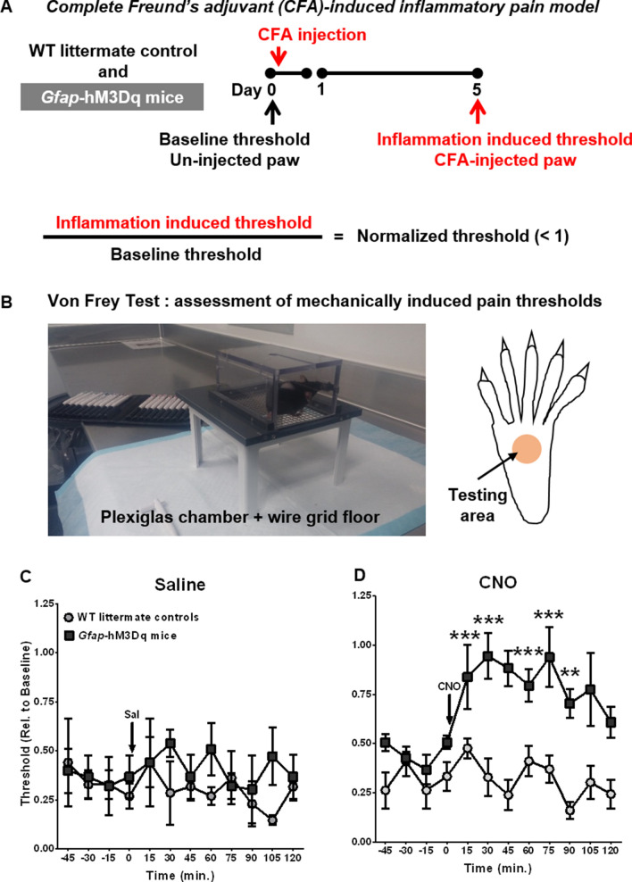
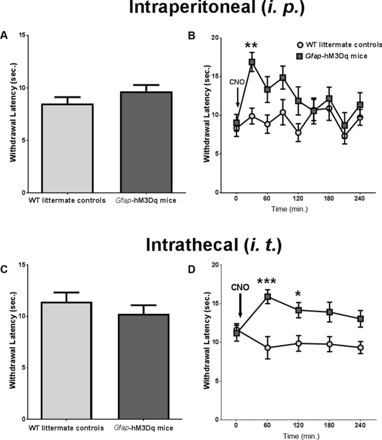
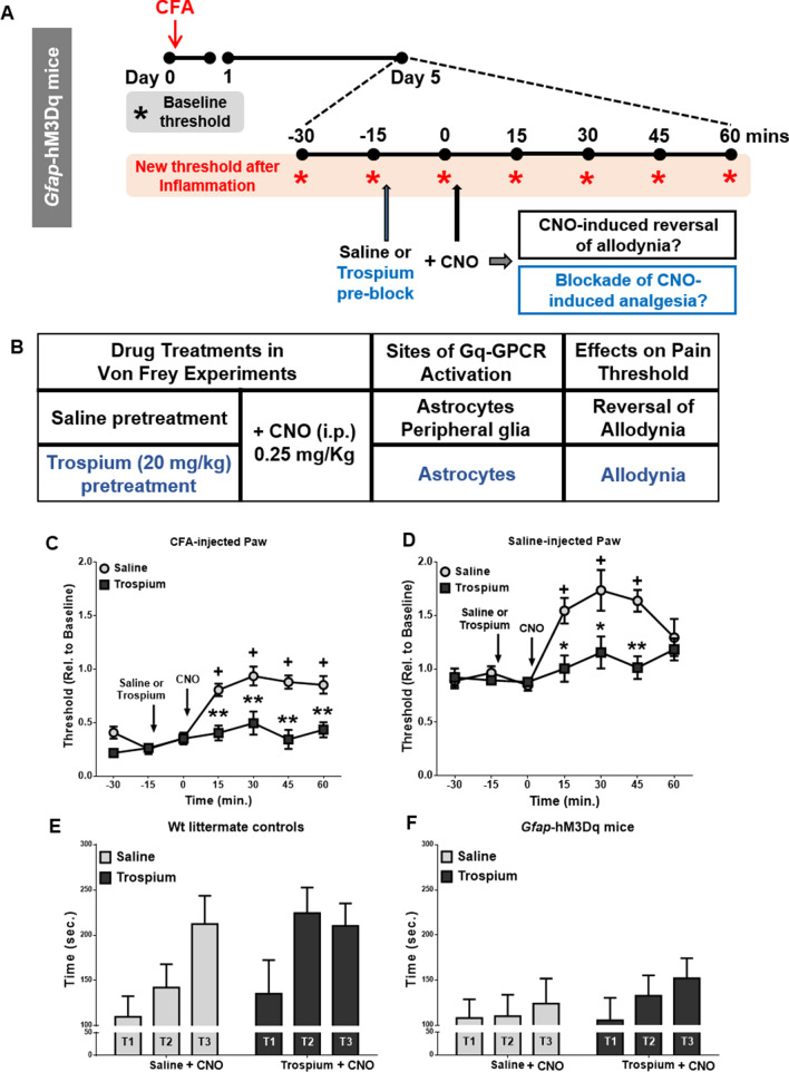
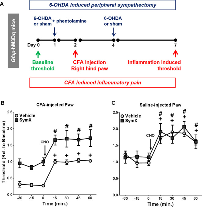
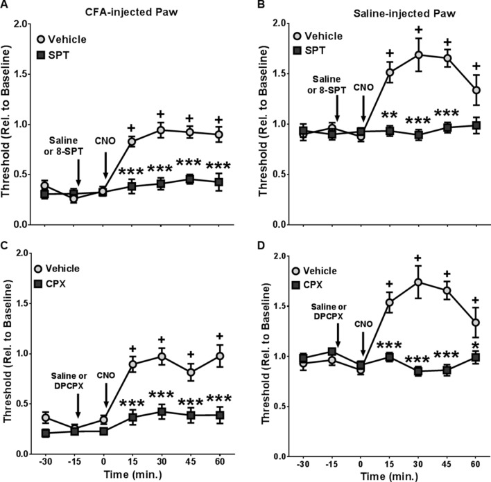
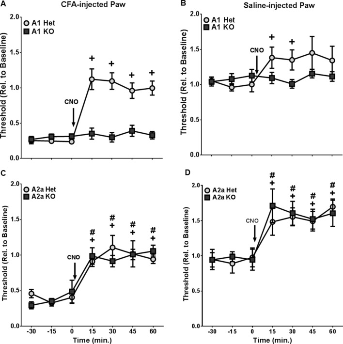
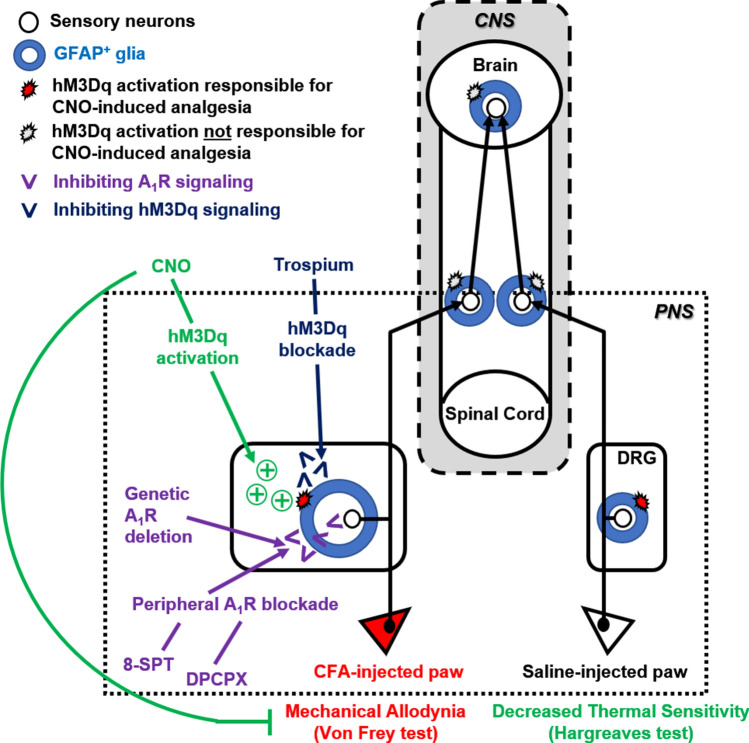
Glial fibrillary acidic protein expressing (GFAP+) glia modulate nociceptive neuronal activity in both the peripheral nervous system (PNS) and the central nervous system (CNS). Resident GFAP+ glia in dorsal root ganglia (DRG) known as satellite glial cells (SGCs) potentiate neuronal activity by releasing pro-inflammatory cytokines and neuroactive compounds. In this study, we tested the hypothesis that SGC Gq-coupled receptor (Gq-GPCR) signaling modulates pain sensitivity in vivo using Gfap-hM3Dq mice. Complete Freund's adjuvant (CFA) was used to induce inflammatory pain, and mechanical sensitivity and thermal sensitivity were used to assess the neuromodulatory effect of glial Gq-GPCR activation in awake mice. Pharmacogenetic activation of Gq-GPCR signaling in sensory SGCs decreased heat-induced nociceptive responses and reversed inflammation-induced mechanical allodynia via peripheral adenosine A1 receptor activation. These data reveal a previously unexplored role of sensory SGCs in decreasing afferent excitability. The identified molecular mechanism underlying the analgesic role of SGCs offers new approaches for reversing peripheral nociceptive sensitization.


Conflict of interest statement

The authors declare no competing interests.

Figures

Figure 1
Figure 1
CNO-induced GFAP+ glial Gq-GPCR activation alleviated CFA-induced mechanical allodynia in vivo. (A) Schematic timeline of CFA-induced inflammatory pain animal model; (B) Von Frey apparatus for assessing mechanically induced pain thresholds. (C) CFA injection into hind paw induced comparable levels of mechanical allodynia in both Gfap-hM3Dq mice and WT littermate controls (N = 3 per genotype). (D) 0.25 mg/kg CNO i. p. administration largely reversed CFA-induced mechanical allodynia in CFA-injected paws in Gfap-hM3Dq mice, but not in WT littermate controls (N = 5 per genotype); **p < 0.01; ***p < 0.001 compared to pre-CNO. (B) was taken by Alison Xie; (C) and (D) were prepared using Graphpad Prism 7.04.
Figure 2
Figure 2
CNO-induced GFAP+ glial Gq-GPCR activation decreased heat sensitivity in the hind paws of naive mice. (A) Baseline heat sensitivity without any drug administration of animals used in (B). Gfap-hM3Dq mice exhibited similar heat sensitivity compared to their WT littermate controls in Hargreaves test (N = 9 control, N = 10 Gfap-hM3Dq). (B) 0.25 mg/kg CNO i. p. administration significantly increased paw withdrawal frequency at 30 min after CNO delivery (N = 9 control, N = 10 Gfap-hM3Dq). (C) Baseline heat sensitivity without any drug administration of animals used in (D). Gfap-hM3Dq mice exhibited similar heat sensitivity compared to their WT littermate controls (N = 10 control, N = 17 Gfap-hM3Dq). (D) Intrathecal administration (i. t.) of 0.5 mg/kg CNO significantly increased paw withdrawal latency up to 2 h (N = 10 control, N = 17 Gfap-hM3Dq). In both (B) and (D), *p < 0.05; **p < 0.01; ***p < 0.001 compared to pre-CNO. Figures were prepared using Graphpad Prism 7.04.
Figure 3
Figure 3
CNO-induced analgesia in Gfap-hM3Dq mice was due to Gq-GPCR activation in peripheral GFAP+ glia and not due to CNO-induced decreases in motor activity. (A) Schematic timeline of peripheral hM3Dq blockade experiments Gfap-hM3Dq mice were subjected to CFA-induced inflammatory pain model as in Fig. 1. Von Frey tests were used to assess both baseline and inflammation induced mechanical pain threshold in both CFA-injected and saline-injected hind paws (N = 21 mice). (B) A summary of peripheral Gq-GPCR activation blockade in Gfap-hM3Dq mice with saline or Trospium pretreatment. (C,D) Trospium pretreatment prevented CNO-induced increases of pain threshold in both CFA-injected (C) and saline-injected (D) hind paws in Gfap-hM3Dq mice (N = 11 saline pretreated, N = 10 trospium pretreated). (E) 0.25 mg/kg CNO i. p. administration had no effect on rotarod performance (three trials, T1, T2, and T3) in WT littermate control mice, nor did pretreatment with trospium (N = 6 per group). (F) CNO led to decreased rotarod performance in Gfap-hM3Dq mice. Trospium pretreatment did not alter CNO-induced decreases in rotarod performance (N = 5 per group). +p < 0.01 compared to baseline in saline-treated mice; *p < 0.01, **p < 0.001 compared to saline-treated mice. (CF) Were prepared using Graphpad Prism 7.04.
Figure 4
Figure 4
Peripheral chemical sympathectomy reversed inflammation induced mechanical allodynia in CFA-injected hind paws in Gfap-hM3Dq mice, while CNO induced additional analgesia effects in both CFA-injected and saline-injected hind paws. (A) Experimental timeline of 6-OHDA induced peripheral chemical sympathectomy and CFA-induced inflammatory pain in Gfap-hM3Dq mice (N = 6 vehicle injection, N = 5 6-OHDA injection). (B) Peripheral sympathectomy blocked CFA-induced mechanical allodynia in Gfap-hM3Dq mice. CNO-induced analgesia was observed in CFA-injected hind paws in both the vehicle- and 6-OHDA-injected groups. (C) Sympathectomy does not affect either the baseline pain threshold or CNO-induced analgesia in saline-injected hind paw. +p < 0.01 compared to baseline in vehicle treated mice; #p < 0.01 compared to baseline in sympathectomized mice. (B,C) Were prepared using Graphpad Prism 7.04.
Figure 5
Figure 5
Pharmacological blockade of peripheral A1 Adenosine receptors prevented CNO-induced analgesia in Gfap-hM3Dq mice. Peripheral administration of SPT, a blood brain barrier impermeable inhibitor of adenosine receptors ablated CNO-induced analgesia in CFA-injected (A) and saline-injected (B) hind paws (N = 13 saline treated, N = 5 SPT treated). A1 adenosine receptor blocker CPX also prevented CNO-induced analgesia in CFA-injected (C) and saline-injected (D) hind paws (N = 13 saline treated, N = 12 CPX treated). +p < 0.01 compared to baseline in saline-treated animals; *p < 0.05, **p < 0.01, ***p < 0.001 compared to saline-treated animals. Figures were prepared using Graphpad Prism 7.04.
Figure 6
Figure 6
Gfap-hM3Dq mice lacking A1R, but not those lacking A2AR failed to exhibit CNO-induced analgesia. CNO-induced analgesia was observed in A1 heterozygous (Het) but not in A1 knockout (KO) mice in both CFA-injected (A) and saline-injected (B) hind paws (N = 9 A1 Het mice, and N = 13 A1 KO mice). CNO-induced analgesia remained intact in both A2A heterozygous and in A2A knockout mice in both the CFA-injected (C) and saline-injected (D) hind paws (N = 7 per group). +p < 0.01 compared to baseline in heterozygous mice; #p < 0.01 compared to baseline in knockout mice. Figures were prepared using Graphpad Prism 7.04.
Figure 7
Figure 7
Summary of experimental strategies and approaches. Gfap-hM3Dq mice express hM3Dq, a Gq-coupled engineered GPCR in GFAP+ glial cells throughout the nervous system. The ligand of hM3Dq, CNO effectively induces hind paw mechanical analgesia (Fig. 1) and decreases thermal sensitivity (Fig. 2) in GFAP-hM3Dq mice. To distinguish the potential analgesic effect of CNS astrocytes vs PNS GFAP+ glia following their Gq-GPCR activation, a peripheral acting blocker of muscarinic receptors, Trospium chloride, was administrated to block CNO-induced and hM3Dq-mediated Gq-GPCR activation in peripheral glia. Astrocytic Gq-GPCR activation alone did not lead to significant changes in hind paw mechanical sensitivity (Fig. 3), which suggested that peripheral GFAP+ glial Gq-GPCR activation is responsible for CNO-induced hind paw analgesia. The glial-induced analgesic effect was independent from inflammation and not affected by peripheral sympathectomy (Fig. 4), suggesting that peripheral glial Gq-GPCR activation directly modulate sensory neuronal activity and/or afferent excitability. To further test this hypothesis, peripheral acting and selective adenosine receptor A1 adenosine receptor antagonists were administrated prior to CNO in GFAP-hM3Dq mice. Blockade of peripheral A1R activation completely abolished peripheral GFAP+ glial activation induced mechanical analgesia (Fig. 5); similar results were repeated using A1R KO mice that also express hM3Dq in GFAP+ glia (Fig. 6). These experiments strongly suggested that peripheral glial Gq-GPCR activation decreases hind paw mechanical sensitivity via peripheral activation of A1R.

Similar articles

Cited by

References

    1. Ji R, Andrea N, Huh Y, Terrando N, Maixner W. Neuroinflammation and central sensitization in chronic and generalized pain. Anestesiología. 2018;19:343–366. - PMC - PubMed
    1. Gold MS, Gebhart GF. Nociceptor sensitization in pain pathogenesis Michael. Mater. Med. 2010;16:1248–1257. - PMC - PubMed
    1. Ji R, Berta T, Nedergaard M. Glia and pain: Is chronic pain a gliopathy? Pain. 2013;154:S10–S28. - PMC - PubMed
    1. Gosselin R, Suter MR, Ji R. Glial cells and chronic pain. Neuroscientist. 2011;16:519–531. - PMC - PubMed
    1. Honore P, et al. Murine models of inflammatory, neuropathic and cancer pain each generates a unique set of neurochemical changes in the spinal cord and sensory neurons. Neuroscience. 2000;98:585–598. - PubMed

Publication types

MeSH terms

LinkOut - more resources