. 2020 Oct;1(6):e245-e253.
doi: 10.1016/S2666-5247(20)30115-4. Epub 2020 Aug 20.

Histopathological findings and viral tropism in UK patients with severe fatal COVID-19: a post-mortem study

Affiliations

Histopathological findings and viral tropism in UK patients with severe fatal COVID-19: a post-mortem study

Brian Hanley et al. Lancet Microbe. 2020 Oct.

Abstract

Background: Severe COVID-19 has a high mortality rate. Comprehensive pathological descriptions of COVID-19 are scarce and limited in scope. We aimed to describe the histopathological findings and viral tropism in patients who died of severe COVID-19.

Methods: In this case series, patients were considered eligible if they were older than 18 years, with premortem diagnosis of severe acute respiratory syndrome coronavirus 2 infection and COVID-19 listed clinically as the direct cause of death. Between March 1 and April 30, 2020, full post-mortem examinations were done on nine patients with confirmed COVID-19, including sampling of all major organs. A limited autopsy was done on one additional patient. Histochemical and immunohistochemical analyses were done, and histopathological findings were reported by subspecialist pathologists. Viral quantitative RT-PCR analysis was done on tissue samples from a subset of patients.

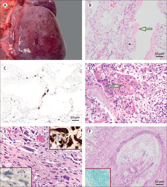
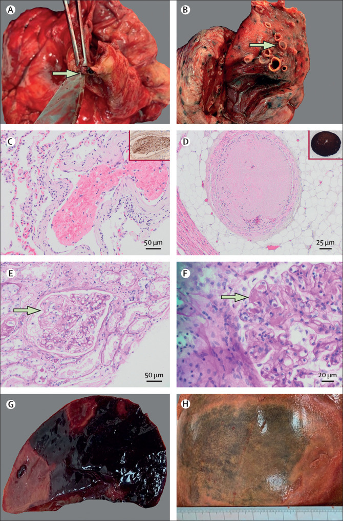
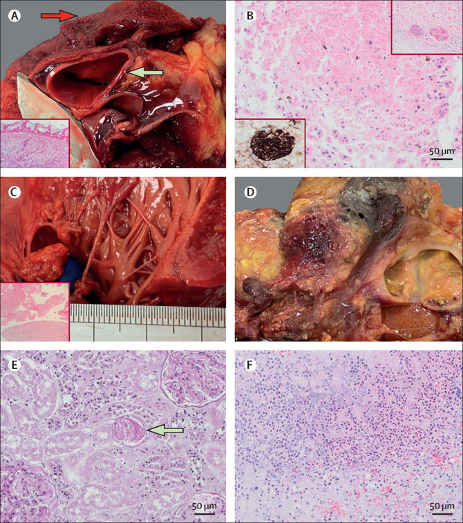
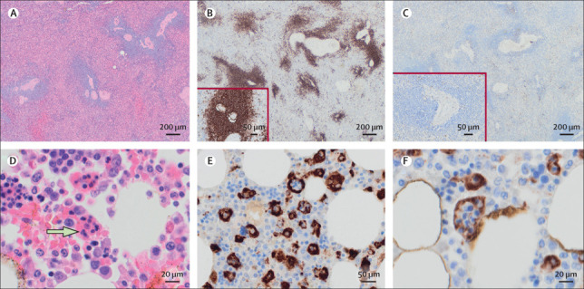
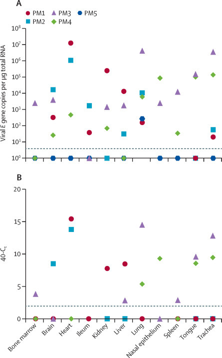
Findings: The median age at death of our cohort of ten patients was 73 years (IQR 52-79). Thrombotic features were observed in at least one major organ in all full autopsies, predominantly in the lung (eight [89%] of nine patients), heart (five [56%]), and kidney (four [44%]). Diffuse alveolar damage was the most consistent lung finding (all ten patients); however, organisation was noted in patients with a longer clinical course. We documented lymphocyte depletion (particularly CD8-positive T cells) in haematological organs and haemophagocytosis. Evidence of acute tubular injury was noted in all nine patients examined. Major unexpected findings were acute pancreatitis (two [22%] of nine patients), adrenal micro-infarction (three [33%]), pericarditis (two [22%]), disseminated mucormycosis (one [10%] of ten patients), aortic dissection (one [11%] of nine patients), and marantic endocarditis (one [11%]). Viral genomes were detected outside of the respiratory tract in four of five patients. The presence of subgenomic viral RNA transcripts provided evidence of active viral replication outside the respiratory tract in three of five patients.

Interpretation: Our series supports clinical data showing that the four dominant interrelated pathological processes in severe COVID-19 are diffuse alveolar damage, thrombosis, haemophagocytosis, and immune cell depletion. Additionally, we report here several novel autopsy findings including pancreatitis, pericarditis, adrenal micro-infarction, secondary disseminated mucormycosis, and brain microglial activation, which require additional investigation to understand their role in COVID-19.

Funding: Imperial Biomedical Research Centre, Wellcome Trust, Biotechnology and Biological Sciences Research Council.


Figures

Figure 1
Figure 1
Pulmonary pathological findings in patients with COVID-19 (A) Macroscopic subpleural petechial haemorrhage in a 24-year-old man (PM6). (B) Hyaline membranes indicative of exudative phase diffuse alveolar damage in a 79-year-old woman (PM9) at 20x magnification. (C) CD61 immunohistochemical staining indicating platelet-rich microthrombosis in alveolar capillaries (PM6). (D) Squamous metaplasia in a 61-year-old man (PM1) with exudative phase diffuse alveolar damage at 40× magnification. (E) Interstitial multinucleated giant cells in a 79-year-old man (PM7) with organising phase diffuse alveolar damage at 40× magnification; the top right insert is of multinucleated giant cells showing positive CD68 staining, indicative of macrophage lineage; the bottom left insert shows absence of staining for cytokeratins. (F) Periodic acid Schiff staining indicating wide, irregular, aseptate, and ribbon-like hyphae with open-angle branching and a vasculocentric pattern indicative of mucormycosis in a 22-year-old man (PM5); the insert is a Grocott silver stain highlighting mucormycosis at 20x magnification.
Figure 2
Figure 2
Thrombotic features identified at autopsy in patients with COVID-19 (A) Macroscopic right coronary artery thrombosis (arrow) in a 61-year-old man (PM1) with exudative phase diffuse alveolar damage. (B) Macroscopic pulmonary thromboembolism (arrow) in a 97-year-old man (PM8). (C) Thrombus in the lung of a 79-year-old woman (PM9) on haematoxylin and eosin staining at 20X magnification; the insert shows CD61 immunohistochemistry indicating moderate staining for platelets. (D) Platelet-rich thrombus in the medium-sized vessels surrounding the heart in a 61-year-old man (PM1); the insert shows strong CD61 staining for platelets. Periodic acid Schiff staining showing a glomerular microaneurysm (arrow, E) and microthrombi within glomerular capillary loops (arrow, F, at 40X magnification) indicative of thrombotic microangiopathy in a 97-year-old man (PM8). Macroscopic splenic (G) and hepatic (H) infarction in a 22-year old man (PM5).
Figure 3
Figure 3
Other notable autopsy findings in patients with COVID-19 (A) Contained aortic dissection (green arrow) and fibrinous pericarditis (red arrow) in a 22-year-old man (PM5); insert is a haematoxylin and eosin stain image of the pericardium showing fibrinous pericarditis (10X magnification). (B) Adrenocortical micro-infarcts in a 79-year-old woman (PM9) with re-endothelialising thrombus in small adrenal vessels highlighted by CD34 (insert, bottom left) and haematoxylin and eosin (insert, top right). Marantic endocarditis (C) highlight with haematoxylin and eosin staining (bottom left at 10x magnification) and necrotising, haemorrhagic pancreatitis (D) in a 22-year-old man (PM5) with COVID-19 and a secondary fungal lung infection. (E) Periodic acid Schiff staining showing a granular cast (arrow) indicative of acute tubular injury in a 24-year-old man (PM6; 20X magnification). (F) Microscopic acute pancreatitis on haematoxylin and eosin staining in a 97-year-old man (PM8; 20X magnification).
Figure 4
Figure 4
Pathological findings in haematological organs in patients with COVID-19 T-cell depletion in the spleen of a 79-year-old woman (PM9) with COVID-19: haematoxylin and eosin staining of the spleen at 10X magnification (A); CD20 staining of spleen indicating presence of B cells (B; 10X magnification), with the insert showing the same region at higher power (20X magnification); and CD3 staining of spleen indicating depletion of T cells (C; 10X magnification), with the insert showing the same region at higher power (20X magnification). Bone marrow phagocytosis in a 97-year-old man (PM8) with COVID-19: haematoxylin and eosin staining of a well preserved bone marrow, with an arrow indicating presence of phagocytosis (D; 40X magnification); and CD68-PGM1 staining of bone marrow indicating presence of phagocytosis (20X [E] and 40x [F] magnification).
Figure 5
Figure 5
Tissue tropism of SARS-CoV-2 in post-mortem samples Fresh tissues were collected from a subset of post-mortem examinations and viral load quantified by use of qRT-PCR targeting the viral E gene (A). Detection of viral RNA was verified by use of qRT-PCR against the viral polymerase gene (data not shown). Tissues were additionally tested for subgenomic viral RNA transcripts (B). Dotted lines indicate the limit of detection as ascertained by negative control. Data are included for a 61-year-old man (PM1), a 64-year-old man (PM2), a 69-year-old woman (PM3), a 78-year-old man (PM4), and a 22-year-old man (PM5). qRT-PCR=quantitative RT-PCR. SARS-CoV-2=severe acute respiratory syndrome coronavirus 2.

References

    1. European Centre for Disease Prevention and Control Threats and outbreaks: COVID-19. 2020. https://www.ecdc.europa.eu/en/novel-coronavirus-china
    1. Huang C, Wang Y, Li X. Clinical features of patients infected with 2019 novel coronavirus in Wuhan, China. Lancet. 2020;395:497–506. - PMC - PubMed
    1. Hanley B, Lucas SB, Youd E, Swift B, Osborn M. Autopsy in suspected COVID-19 cases. J Clin Pathol. 2020;73:239–242. - PubMed
    1. Ferrario CM, Jessup J, Chappell MC. Effect of angiotensin-converting enzyme inhibition and angiotensin II receptor blockers on cardiac angiotensin-converting enzyme 2. Circulation. 2005;111:2605–2610. - PubMed
    1. Hoffmann M, Kleine-Weber H, Schroeder S. SARS-CoV-2 cell entry depends on ACE2 and TMPRSS2 and is blocked by a clinically proven protease inhibitor. Cell. 2020;181:271–280. - PMC - PubMed

Publication types