

doi: 10.1007/s40477-020-00526-y.
Epub 2020 Aug 25.
Lung ultrasound artifacts in COVID-19 patients
Affiliations
- PMID: 32844375
- PMCID: PMC7446742
- DOI: 10.1007/s40477-020-00526-y
Item in Clipboard
Lung ultrasound artifacts in COVID-19 patients
J Ultrasound.
2022 Jun.
Abstract
Lung ultrasound is an essential tool in critical care, made more so by the enhanced precautions associated with the Covid-19 pandemic. Here we describe 2 cases of multiple, small shred signs seen on ultrasound of Covid-19 patients.
Keywords: COVID-19; Lung ultrasound; Shred sign.
© 2020. Società Italiana di Ultrasonologia in Medicina e Biologia (SIUMB).
Figures
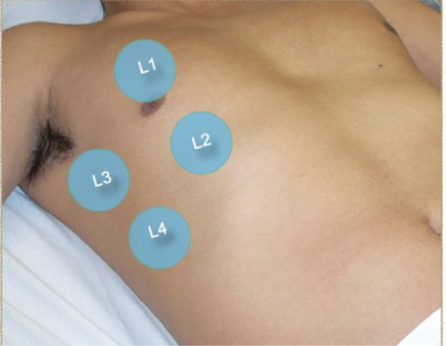
Lung zones in a supine position. L1 Upper anterior lung zone. L2 Lower anterior lung zone. L3 Upper lateral lung zone. L4 Lower lateral lung zone

a, a’ Patient A and B chest X-ray prior to lung ultrasound with bibasilar infiltrates. b, b’ Pleural lines for patients A and B with multiple small shred signs (arrows). c, c’ illustrating the grainy appearance of the pleural line resembling pebbles in patients A and B, identified by the arrows. d Showing skip areas of normal to mildly thickened pleural line with multiple B-Lines (arrow heads)

a, a’ Patient A and B chest X-ray prior to lung ultrasound with bibasilar infiltrates. b, b’ Pleural lines for patients A and B with multiple small shred signs (arrows). c, c’ illustrating the grainy appearance of the pleural line resembling pebbles in patients A and B, identified by the arrows. d Showing skip areas of normal to mildly thickened pleural line with multiple B-Lines (arrow heads)

Illustration of the pathophysiology of the different ultrasound signs including the pebbles resembling artifacts. Note areas of normal lung parenchyma and air-filled alveoli with poor transmission to ultrasound, reflecting and scattering the waves (Arrows) as bright pebbles like artifacts. Also, areas of lung tissue with early buildup and accumulation of alveolar secretions, mucous and inflammatory cells, leading to transmission of the ultrasound beam and its refraction to deeper tissues (arrow heads). Thickening of the interstitial layer with fluid accumulation can lead to the reverberation of the ultrasound beam between the pleural line and the interlobular septa, leading to the development of the comet tail, or B Lines on the ultrasound screen
References
-
- Tierney DM et al. (2020) Comparative performance of pulmonary ultrasound, chest radiograph, and CT among patients with acute respiratory failure. Crit Care Med 48(2):151–157. ISSN 1530–0293. Disponível em https://www.ncbi.nlm.nih.gov/pubmed/31939782. - PubMed
-
- https://www.covid19treatmentguidelines.nih.gov/overview/. NIH Coronavirus disease 2019 (COVID-19) treatment guidelines. NIH.gov, 2020. - PubMed
-
- Center JHU, A. M. C. R. Johns Hopkins University and Medicine/Coronavirus Resource Center: https://coronavirus.jhu.edu 2020.
-
- Buonsenso D et al. Point-of-care lung ultrasound findings in novel coronavirus disease-19 pnemoniae: a case report and potential applications during COVID-19 outbreak. Eur Rev Med Pharmacol Sci 24(5):2776–2780, 03 2020. ISSN 2284–0729. Disponível em: https://www.ncbi.nlm.nih.gov/pubmed/32196627. - PubMed
-
- Soldati G et al. Proposal for international standardization of the use of lung ultrasound for patients with COVID-19: a simple, quantitative, reproducible method. J Ultrasound Med 39(7):1413–1419, Jul 2020. ISSN 1550–9613. Disponível em: https://www.ncbi.nlm.nih.gov/pubmed/32227492. - PMC - PubMed
MeSH terms
LinkOut - more resources
Full Text Sources
Medical
