. 2020 Nov 12;136(20):2334-2345.
doi: 10.1182/blood.2019004332.

The JAK-STAT pathway regulates CD38 on myeloma cells in the bone marrow microenvironment: therapeutic implications

Affiliations

The JAK-STAT pathway regulates CD38 on myeloma cells in the bone marrow microenvironment: therapeutic implications

Daisuke Ogiya et al. Blood. .

Abstract

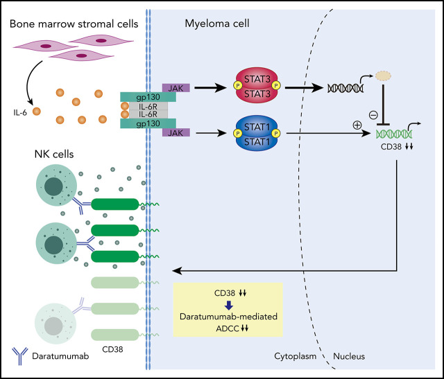
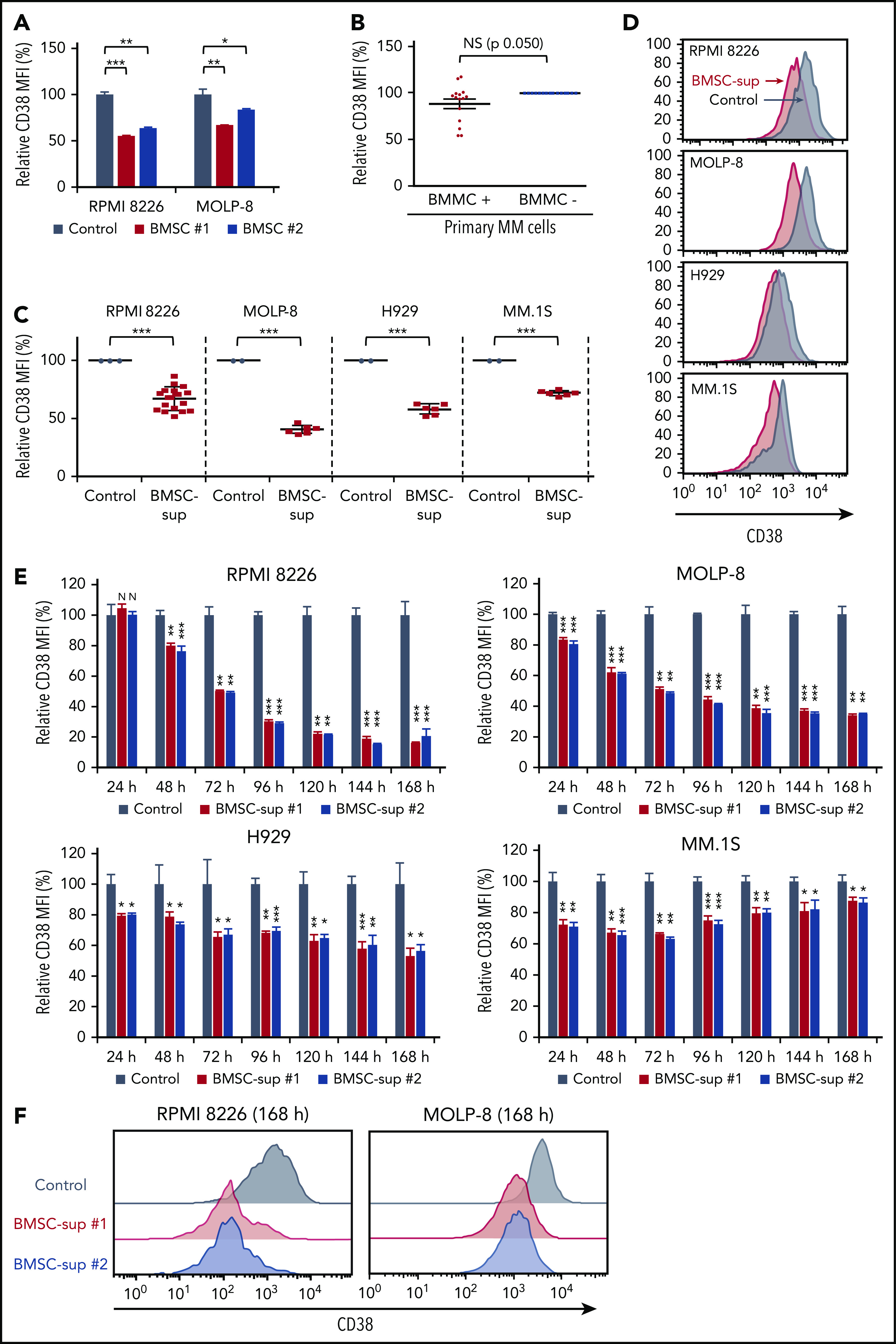
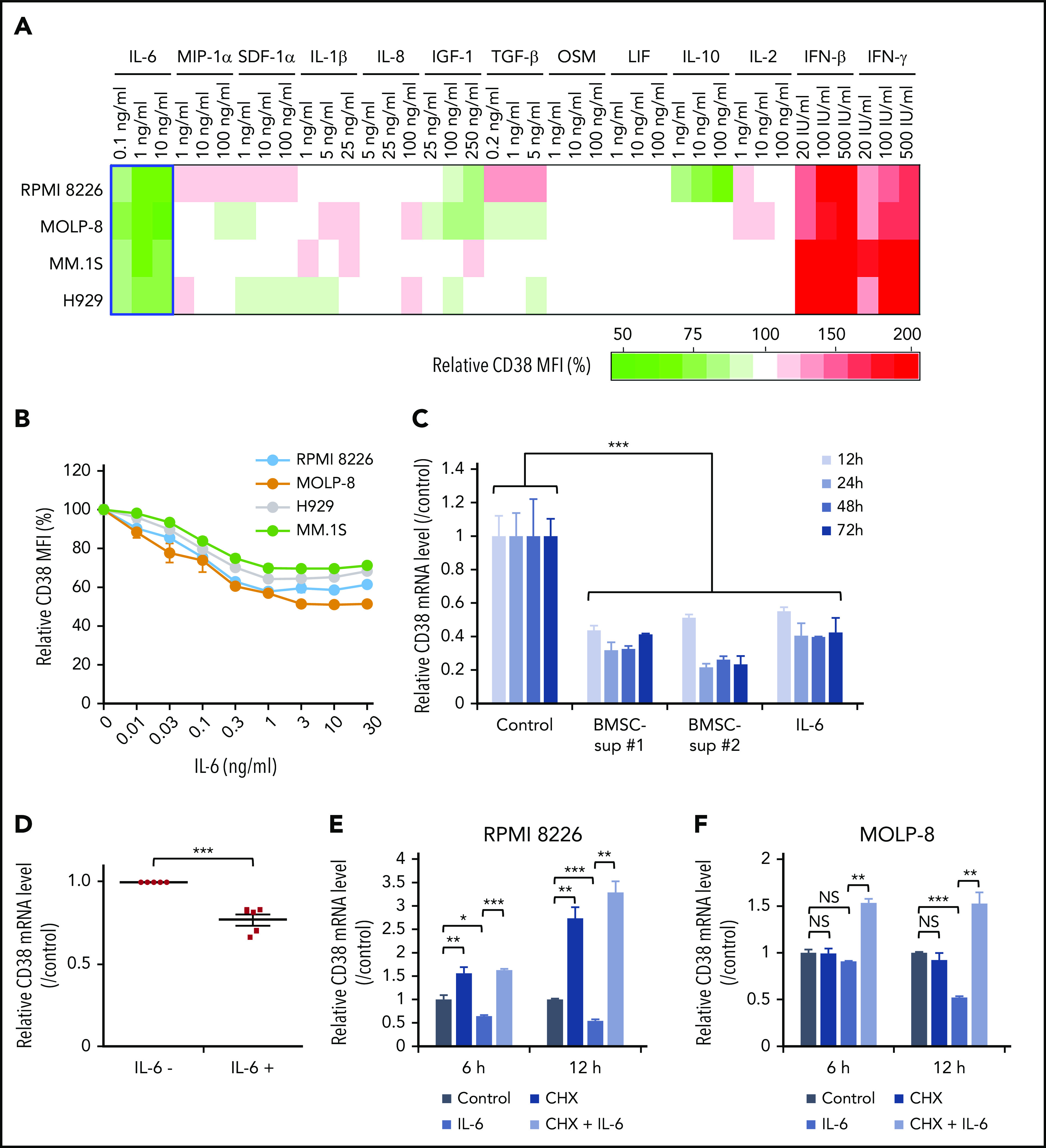
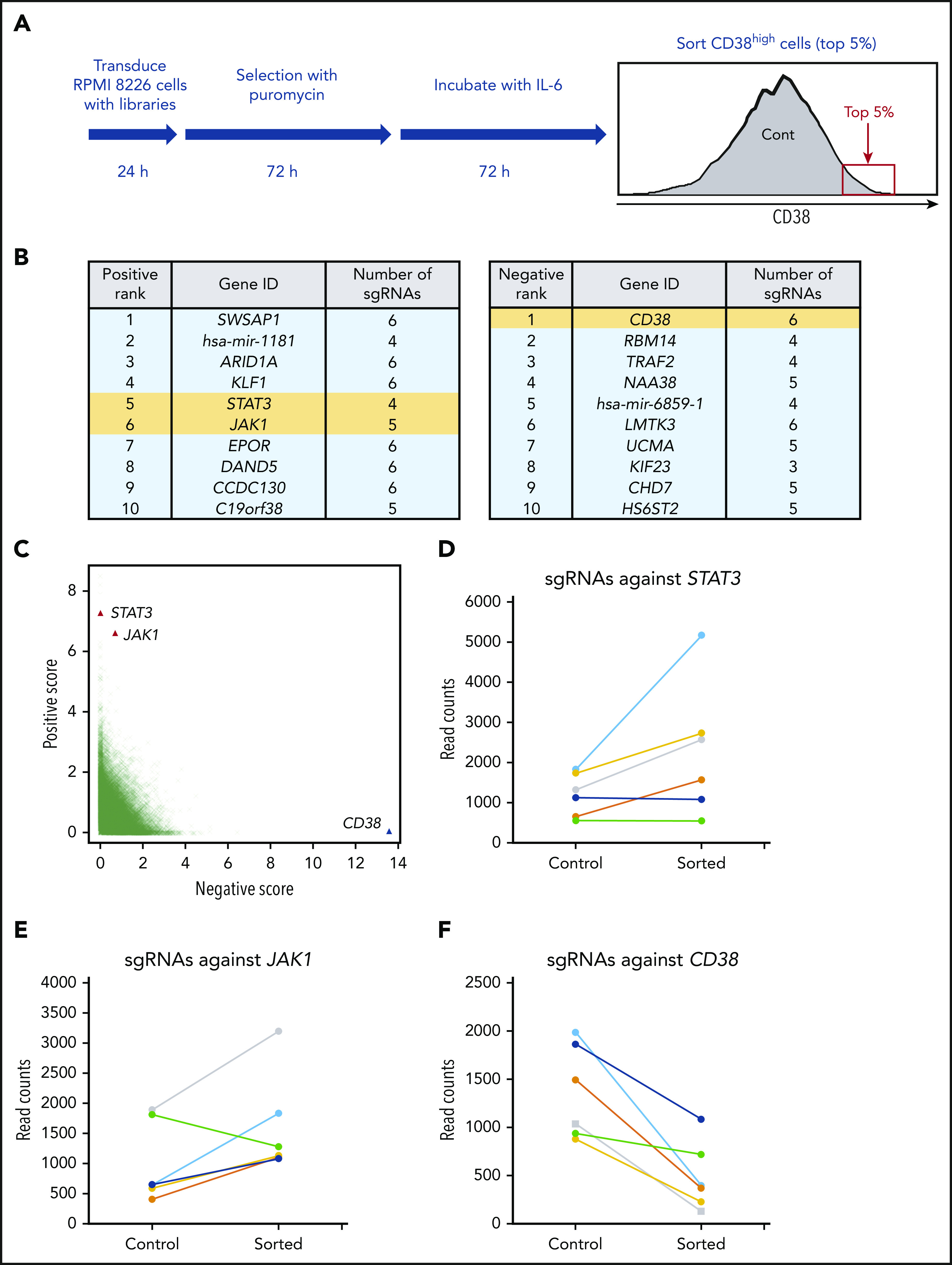
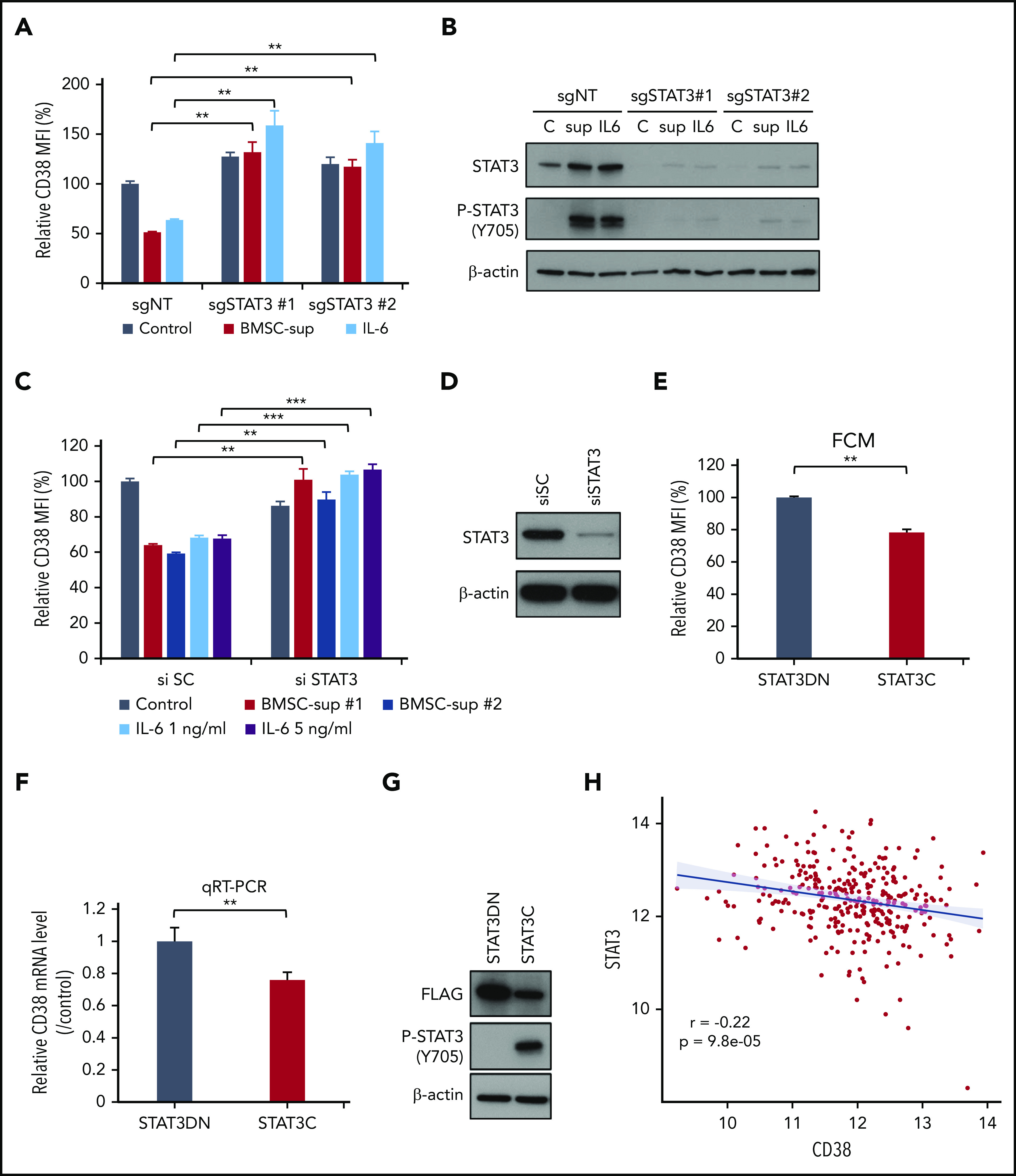
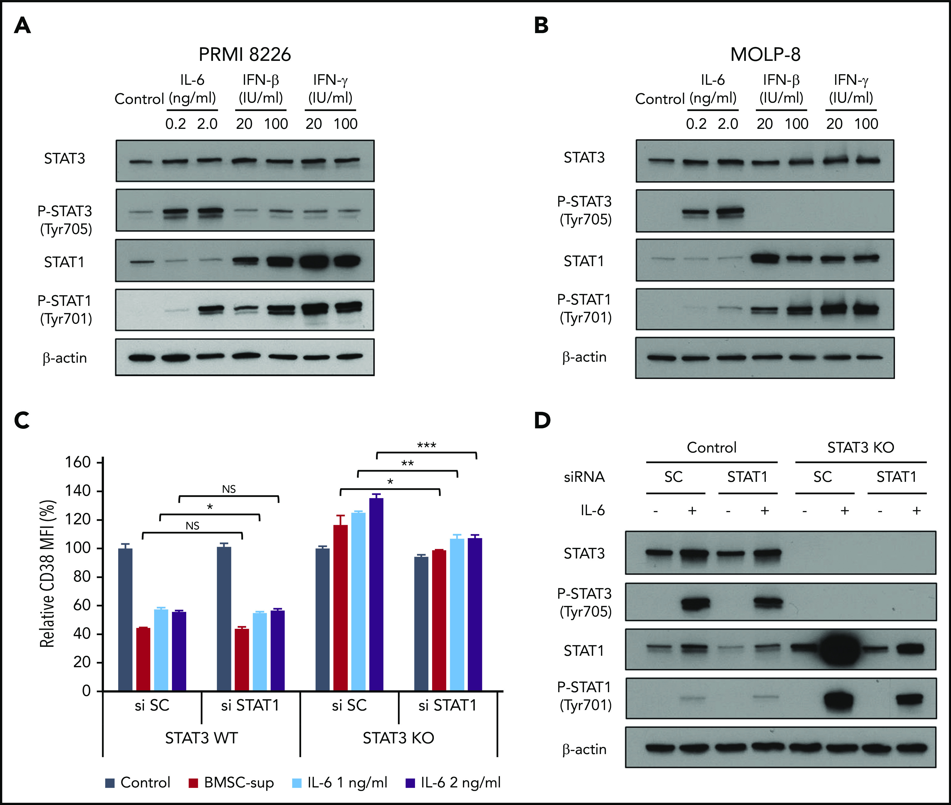
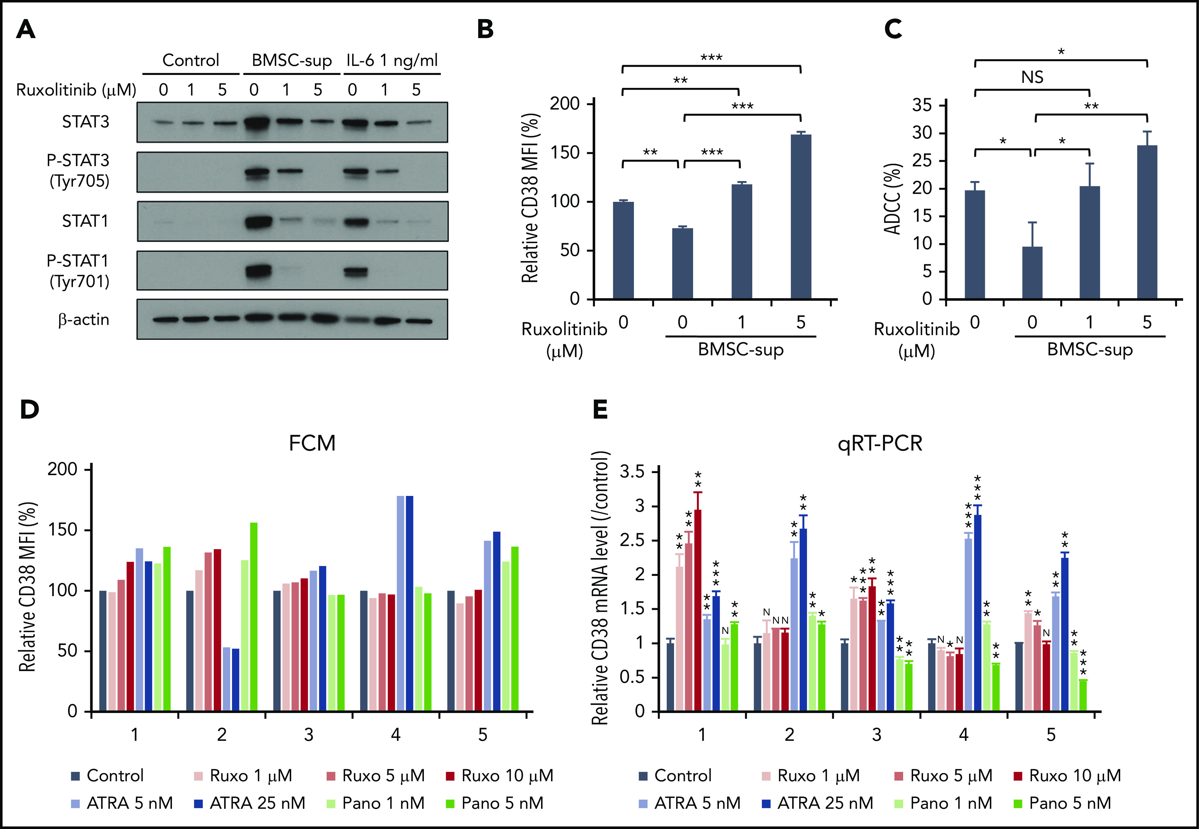
Anti-CD38 monoclonal antibody (MoAb) treatments including daratumumab (DARA) are effective therapies for both newly diagnosed and relapsed multiple myeloma (MM). In this study, we examined the soluble factors that modulate CD38 expression and are associated with sensitivity to DARA-mediated antibody-dependent cellular cytotoxicity (ADCC) in the bone marrow (BM) microenvironment. Importantly, primary BM stromal cell (BMSC) culture supernatant (BMSC-sup) and interleukin-6 (IL-6) downregulated CD38 expression and reduced DARA-mediated ADCC. Both cytokine profiling of the BMSC-sup and genome-scale clustered regularly interspaced short palindromic repeats (CRISPR)-CRISPR-associated protein 9 (Cas9) knockout screening in MM cell lines identified and validated the JAK-STAT3 signaling pathway mediating CD38 downregulation, whereas the JAK-STAT1 pathway mediated CD38 upregulation. STAT3 knockdown abrogated BMSC-sup- and IL-6-induced CD38 downregulation on MM cell lines. We also confirmed that STAT3 and CD38 is negatively correlated in primary MM cells. To assess potential clinical relevance, pharmacological inhibition of the JAK-STAT pathway on BMSC-sup-induced CD38 downregulation was further examined. JAK inhibitor ruxolitinib inhibited STAT3 phosphorylation in MM cell lines, upregulated CD38 expression in MM cell lines and primary patient MM cells, and augmented DARA-mediated ADCC against MM cell lines. Taken together, our results suggest that CD38 expression on MM cells in the BM microenvironment is regulated by both STAT1 (positively) and STAT3 (negatively), and that inhibition of the JAK-STAT3 pathway represents a novel therapeutic option to enhance CD38 expression and anti-CD38 MoAb-mediated MM cytotoxicity.


Conflict of interest statement

Conflict-of-interest disclosure: K.C.A. serves on advisory boards to Celgene, Millennium-Takeda, Janssen, Sanofi-Aventis, Bristol Myers Squibb, Gilead, Precision Biosciences, and Tolero, and is a scientific founder of OncoPep and C4 Therapeutics. The remaining authors declare no competing financial interests.

Figures

None
Graphical abstract
Figure 1.
Figure 1.
BMSCs and BMSC-sup reduce CD38 expression on MM cells. (A) RPMI 8226 and MOLP-8 cells were cocultured with or without 2 different bone marrow stromal cells (BMSC #1 or #2) for 5 days. After incubation, CD38 expression was measured by flow cytometry, and relative CD38 MFI was calculated in comparison with MFI of control. (B) CD138+ primary MM cells from 15 MM patients were cocultured with or without autologous BMMCs for 5 days. After incubation, CD38 expression on 7-AAD and CD138+ MM cells was measured by flow cytometry, and relative CD38 MFI was calculated in comparison with MFI of MM cells cultured without BMMCs. (C) RPMI 8226, MOLP-8, H929, and MM.1S cells were cultured with control culture medium (control, n = 3 for RPMI 8226, and n = 2 for the other cell lines) or BMSC-sup from different MM patients (n = 17 for RPMI 8226, and n = 6 for the other cell lines) for 72 hours. CD38 expression on 7-AAD cells was measured by flow cytometry, and relative CD38 MFI was calculated in comparison with MFI of control. (D) Representative histograms of CD38 expression of panel C. (E) Relative CD38 MFIs were measured after MM cells were cultured with control medium or 2 different BMSC-sup (BMSC-sup #1 or #2) for 24 to 168 hours. (F) Histograms of CD38 expression in RPMI 8226 and MOLP-8 cells cultured with control medium, BMSC-sup #1 or #2 for 168 hours. Data are shown as mean plus or minus standard error of the mean. *P < .05; **P < .01; ***P < .001.
Figure 2.
Figure 2.
Cytokine profiling shows that IL-6 is the major soluble factor downregulating CD38 expression on MM cells. (A) Four MM cell lines were cultured with control culture medium or culture medium containing the indicated cytokines (IL-6, MIP-1α, SDF-1α, IL-1β, IL-8, IGF-1, TGF-β, OSM, LIF, IL-10, IL-2, IFN-β, IFN-γ) for 72 hours. CD38 expression was measured by flow cytometry after 72-hour incubation, and relative CD38 MFI was calculated in comparison with MFI of control. Heat map was created based on relative CD38 MFI as indicated in supplemental Table 5. (B) Four MM cell lines were treated for 72 hours with IL-6 (0.01-30 ng/mL), and CD38 expression was measured by flow cytometry. (C) RPMI 8226 cells were cultured with control culture media, 2 different BMSC-sup (BMSC-sup #1 or #2), or IL-6 (5 ng/mL) for 12, 24, 48, or 72 hours, and CD38 mRNA level was measured by qRT-PCR. (D) CD138+ primary MM cells separated from BMMCs of 5 MM patients using CD138 magnetic-activated cell separation beads were cultured with or without IL-6 (5 ng/mL), and the CD38 mRNA level was measured by qRT-PCR. (E-F) RPMI 8226 (E) and MOLP-8 (F) cells were cultured with control culture medium or IL-6 (5 ng/mL), in the absence or presence of CHX (10 µg/mL) for 6 hours or 12 hours, and the CD38 mRNA level was measured by qRT-PCR. Data are shown as mean plus or minus standard error of the mean. *P < .05; **P < .01; ***P < .001.
Figure 3.
Figure 3.
Genome-scale CRISPR-Cas9 knockout screening identifies STAT3 as a repressor of CD38. (A) Flowchart of genome-scale CRIPSR-Cas9 screening in RPMI 8226 cells. (B) List of top 10 positive (left panel) and negative (right panel) ranked genes; positive represents sgRNAs enriched in sorted cells compared with control cells, and negative the opposite. STAT3, JAK1, and CD38 are highlighted in yellow. (C) Enrichment of specific sgRNAs in CD38-high fraction. Each dot specifies 1 sgRNA. (D-F) Read counts of sgRNAs against STAT3 (D), JAK1 (E), and CD38 (F) in control and sorted cells.
Figure 4.
Figure 4.
STAT3 negatively regulates CD38 expression. (A-B) RPMI 8226 cells were infected with single-guide (sg) nontarget control (sgNT), sgSTAT3 #1 or #2 expressing lentiviral vector, and cultured with control culture medium (Control), BMSC-sup, or IL-6 (1 ng/mL) for 72 hours. (C-D) RPMI 8226 cells were transfected with scrambled siRNA (si SC) or STAT3 siRNA (si STAT3). These cells were cultured with control culture medium, BMSC-sup, or IL-6 (1 or 5 ng/mL) for 48 hours. After incubation, CD38 expression was measured by flow cytometry (A, C). STAT3 downregulation was confirmed by immunoblotting (B, D). (E-G) RPMI 8226 cells were transfected with the dominant-negative STAT3 (STAT3DN) or constitutively active STAT3 (STAT3C) construct. CD38 protein expression (E) and mRNA level (F) were assessed by flow cytometry (FCM) and qRT-PCR, respectively. (G) Phosphorylated STAT3 (P-STAT3) expression was confirmed by immunoblotting. Relative CD38 MFI was calculated in comparison with MFI of control. Data are shown as mean plus or minus standard error of the mean. *P < .05; **P < .01; ***P < .001. (H) CD38 (x-axis) and STAT3 (y-axis) expression are negatively correlated (r, −0.22; P, 9.8e-05) at diagnosis in 319 newly diagnosed MM patient samples from the IFM/DFCI 2009 study. C, control culture medium; sup, BMSC-sup.
Figure 5.
Figure 5.
Regulation of CD38 by STAT1 and STAT3. (A-B) RPMI 8226 (A) and MOLP-8 (B) cells were cultured with IL-6, IFN-β, or IFN-γ for 6 hours, and then subjected to immunoblotting using indicated antibodies. (C) STAT3 stable knockout (KO) RPMI 8226 cells were transfected with scrambled siRNA (si SC) or STAT1 siRNA (si STAT1), and then cultured with BMSC-sup or IL-6 (1 or 2 ng/mL) for 72 hours. CD38 expression was measured by flow cytometry. (D) STAT3 stable knockout RPMI 8226 cells were transfected with scrambled (SC) siRNA or STAT1 siRNA, cultured in the absence or presence of IL-6 (2 ng/mL) for 6 hours, and then subjected to immunoblotting using indicated antibodies. Relative CD38 MFI was calculated in comparison with MFI of control. Data are shown as mean plus or minus standard error of the mean. *P < .05; **P < .01; ***P < .001.
Figure 6.
Figure 6.
JAK inhibitor ruxolitinib upregulates CD38 expression and enhances DARA-mediated ADCC against MM cells. (A) RPMI 8226 cells were treated with solvent control or ruxolitinib (Ruxo; 1, 5 µM), in the absence or presence of BMSC-sup or IL-6 (1 ng/mL) for 48 hours. Whole-cell lysates were subjected to immunoblotting using indicated antibodies. (B-C) RPMI 8226 cells were treated with solvent control or ruxolitinib (1, 5 µM), in the absence or presence of BMSC-sup for 72 hours, and then subjected to flow cytometric analysis for CD38 expression (B) and DARA-mediated ADCC assay (C). (D) BMMCs of 5 MM patients were treated with solvent control, ruxolitinib (1, 5, 10 µM), ATRA (5, 25 nM), or panobinostat (Pano; 1, 5 nM) for 120 hours. After incubation, CD38 expression on 7-AAD and CD138+ MM cells was measured by flow cytometry. (E) CD138+ MM cells separated from BMMCs of 5 MM patients were treated with solvent control, ruxolitinib (1, 5, 10 µM), ATRA (5, 25 nM), or panobinostat (1, 5 nM) for 120 hours. After incubation, relative CD38 mRNA level was assessed by qRT-PCR. Relative CD38 MFI was calculated in comparison with MFI of control. Data are shown as mean plus or minus standard error of the mean. *P < .05; **P < .01; ***P < .001.

Comment in

  • We need CD38 STAT-JAK.
    Barwick BG, Lonial S. Barwick BG, et al. Blood. 2020 Nov 12;136(20):2246-2248. doi: 10.1182/blood.2020007467. Blood. 2020. PMID: 33180919 No abstract available.

References

    1. Kumar SK, Anderson KC. Immune therapies in multiple myeloma. Clin Cancer Res. 2016;22(22):5453-5460. - PubMed
    1. Iftikhar A, Hassan H, Iftikhar N, et al. Investigational monoclonal antibodies in the treatment of multiple myeloma: a systematic review of agents under clinical development. Antibodies (Basel). 2019;8(2):34. - PMC - PubMed
    1. Facon T, Kumar S, Plesner T, et al. ; MAIA Trial Investigators . Daratumumab plus lenalidomide and dexamethasone for untreated myeloma. N Engl J Med. 2019;380(22):2104-2115. - PMC - PubMed
    1. Moreau P, Attal M, Hulin C, et al. Bortezomib, thalidomide, and dexamethasone with or without daratumumab before and after autologous stem-cell transplantation for newly diagnosed multiple myeloma (CASSIOPEIA): a randomised, open-label, phase 3 study. Lancet. 2019;394(10192):29-38. - PubMed
    1. Mateos MV, Dimopoulos MA, Cavo M, et al. ; ALCYONE Trial Investigators . Daratumumab plus bortezomib, melphalan, and prednisone for untreated myeloma. N Engl J Med. 2018;378(6):518-528. - PubMed

Publication types

MeSH terms