

Differentiating rheumatoid and psoriatic arthritis: a systematic analysis of high-resolution magnetic resonance imaging features-preliminary findings
- PMID: 32845377
- PMCID: PMC7811987
- DOI: 10.1007/s00256-020-03588-5
Differentiating rheumatoid and psoriatic arthritis: a systematic analysis of high-resolution magnetic resonance imaging features-preliminary findings
Abstract
Background: Because of overlapping phenotypical presentations, the diagnostic differentiation of rheumatoid arthritis (RA) and psoriatic arthritis (PsA) remains challenging. Thus, this study aimed to examine the diagnostic value of distinct imaging features obtained by high-resolution 3-T MRI for the diagnostic differentiation.
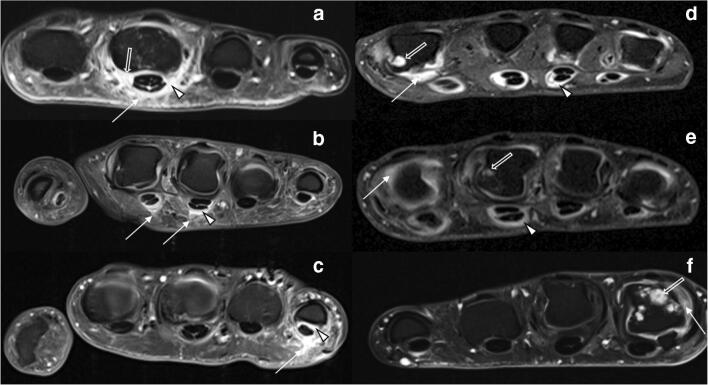
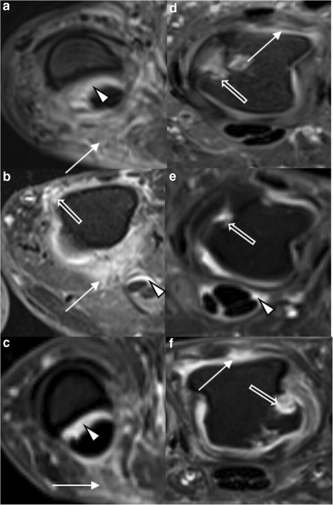
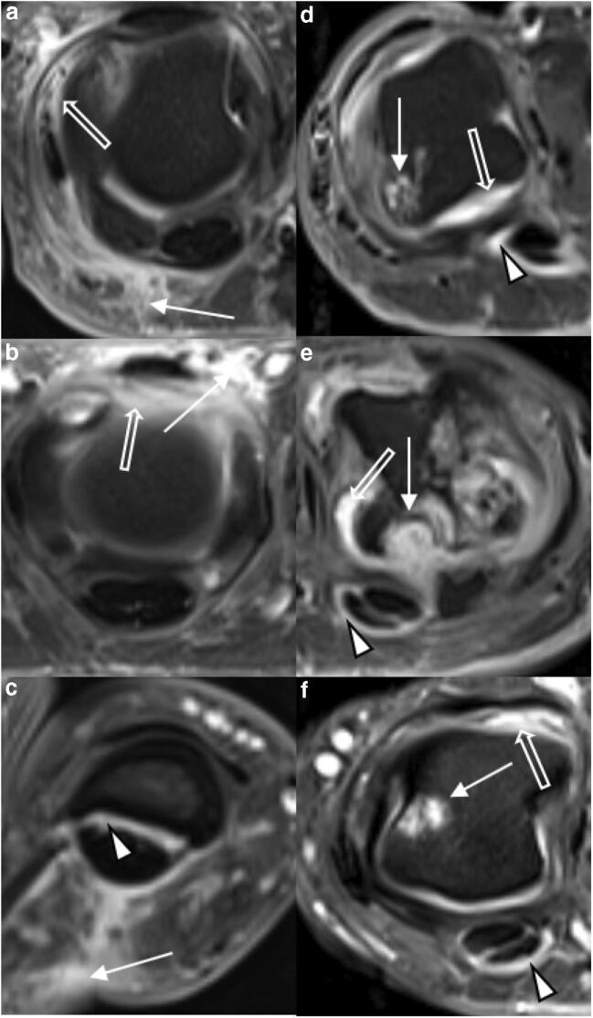
Materials and methods: Seventeen patients with PsA and 28 patients with RA were imaged at high resolution using 3-T MRI scanners and a dedicated 16-channel hand coil. All images were analyzed according to the outcome measures in rheumatology clinical trials' (OMERACT) RAMRIS (Rheumatoid Arthritis Magnetic Resonance Imaging Score) and PsAMRIS (Psoriatic Arthritis Magnetic Resonance Imaging Score) for the presence and intensity of synovitis, flexor tenosynovitis, bone edema, bone erosion, periarticular inflammation, bone proliferation, and joint space narrowing. Next, odds ratios (OR) were calculated to determine the strength of the associations between these imaging features, demographic characteristics, and the outcome RA vs. PsA.
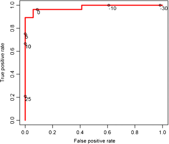
Results: PsA could be differentiated from RA by extracapsular inflammatory changes (PsAMRIS sub-score "periarticular inflammation"), with low odds for the presence of RA (OR of 0.06, p < 0.01) at all metacarpophalangeal (MCP) joints. A prediction model informed by the items that were strongest associated with the presence of RA or PsA demonstrated excellent differentiating capability with an area under the curve of 98.1%.
Conclusion: High-resolution imaging is beneficial for the identification of relevant imaging features that may assist the clinical differentiation of inflammatory conditions of the hand. At the MCP level, extracapsular inflammatory changes were strongly associated with PsA and may consequently allow the imaging differentiation of PsA and RA.
Keywords: MRI; Metacarpophalangeal joint; PsAMRIS; Psoriatic arthritis; RAMRIS; Rheumatoid arthritis.
Conflict of interest statement
The authors declare that they have no competing interests.
Figures
Similar articles
-
Imaging of Peripheral Arthritis: Special Focus on Differences in Inflammatory Lesions Between Rheumatoid Arthritis and Psoriatic Arthritis.Korean J Radiol. 2025 Jun;26(6):569-580. doi: 10.3348/kjr.2025.0036. Epub 2025 Apr 29. Korean J Radiol. 2025. PMID: 40341887 Free PMC article. Review.
-
High-resolution MRI of flexor tendon pulleys using a 16-channel hand coil: disease detection and differentiation of psoriatic and rheumatoid arthritis.Arthritis Res Ther. 2020 Mar 2;22(1):40. doi: 10.1186/s13075-020-2135-0. Arthritis Res Ther. 2020. PMID: 32122392 Free PMC article.
-
Proteoglycan loss in the articular cartilage is associated with severity of joint inflammation in psoriatic arthritis-a compositional magnetic resonance imaging study.Arthritis Res Ther. 2020 May 29;22(1):124. doi: 10.1186/s13075-020-02219-7. Arthritis Res Ther. 2020. PMID: 32471515 Free PMC article.
-
Magnetic resonance imaging-assessed synovial and bone changes in hand and wrist joints of rheumatoid arthritis patients.Korean J Intern Med. 2019 May;34(3):651-659. doi: 10.3904/kjim.2016.271. Epub 2017 Nov 24. Korean J Intern Med. 2019. PMID: 29166759 Free PMC article.
-
The OMERACT MRI in Arthritis Working Group - Update on Status and Future Research Priorities.J Rheumatol. 2015 Dec;42(12):2470-2. doi: 10.3899/jrheum.141248. Epub 2015 Feb 15. J Rheumatol. 2015. PMID: 25684771 Review.
Cited by
-
Comparison of the axillary lymph node between rheumatoid arthritis and psoriatic arthritis with computed tomography.Acta Radiol Open. 2022 Jul 13;11(7):20584601221112616. doi: 10.1177/20584601221112616. eCollection 2022 Jul. Acta Radiol Open. 2022. PMID: 35846390 Free PMC article.
-
Magnetic resonance imaging of rheumatological diseases.Pol J Radiol. 2022 Feb 20;87:e93-e112. doi: 10.5114/pjr.2022.113390. eCollection 2022. Pol J Radiol. 2022. PMID: 35280946 Free PMC article. Review.
-
The COVID-19 Pandemic Heightens Interest in Cytokine Storm Disease and Advances in Machine Learning Diagnosis, Telemedicine, and Primordial Prevention of Rheumatic Diseases.Eur J Rheumatol. 2024 Nov 28;11(4):410-417. doi: 10.5152/eurjrheum.2024.23059. Eur J Rheumatol. 2024. PMID: 39651898 Free PMC article. Review.
-
Imaging of Peripheral Arthritis: Special Focus on Differences in Inflammatory Lesions Between Rheumatoid Arthritis and Psoriatic Arthritis.Korean J Radiol. 2025 Jun;26(6):569-580. doi: 10.3348/kjr.2025.0036. Epub 2025 Apr 29. Korean J Radiol. 2025. PMID: 40341887 Free PMC article. Review.
-
Anatomical analysis of inflammation in hand psoriatic arthritis by Dual-Energy CT Iodine Map.Eur J Radiol Open. 2021 Oct 14;8:100383. doi: 10.1016/j.ejro.2021.100383. eCollection 2021. Eur J Radiol Open. 2021. PMID: 34703848 Free PMC article.
References
-
- Rahman P, Nguyen E, Cheung C, et al. Comparison of radiological severity in psoriatic arthritis and rheumatoid arthritis. J Rheumatol. 2001;28(5):1041–1044. - PubMed
-
- Finzel S, Englbrecht M, Engelke K, et al. A comparative study of periarticular bone lesions in rheumatoid arthritis and psoriatic arthritis. Ann Rheum Dis. 2011;70(1):122–127. - PubMed
-
- Taylor W, Gladman D, Helliwell P, et al. Classification criteria for psoriatic arthritis: development of new criteria from a large international study. Arthritis Rheum. 2006;54(8):2665–2673. - PubMed
MeSH terms
Grants and funding
LinkOut - more resources
Full Text Sources
Medical
Research Materials
Miscellaneous
