. 2020 Oct 15;31(22):2463-2474.
doi: 10.1091/mbc.E19-03-0133. Epub 2020 Aug 26.

Polarized human cholangiocytes release distinct populations of apical and basolateral small extracellular vesicles

Affiliations

Polarized human cholangiocytes release distinct populations of apical and basolateral small extracellular vesicles

Brian A Davies et al. Mol Biol Cell. .

Abstract

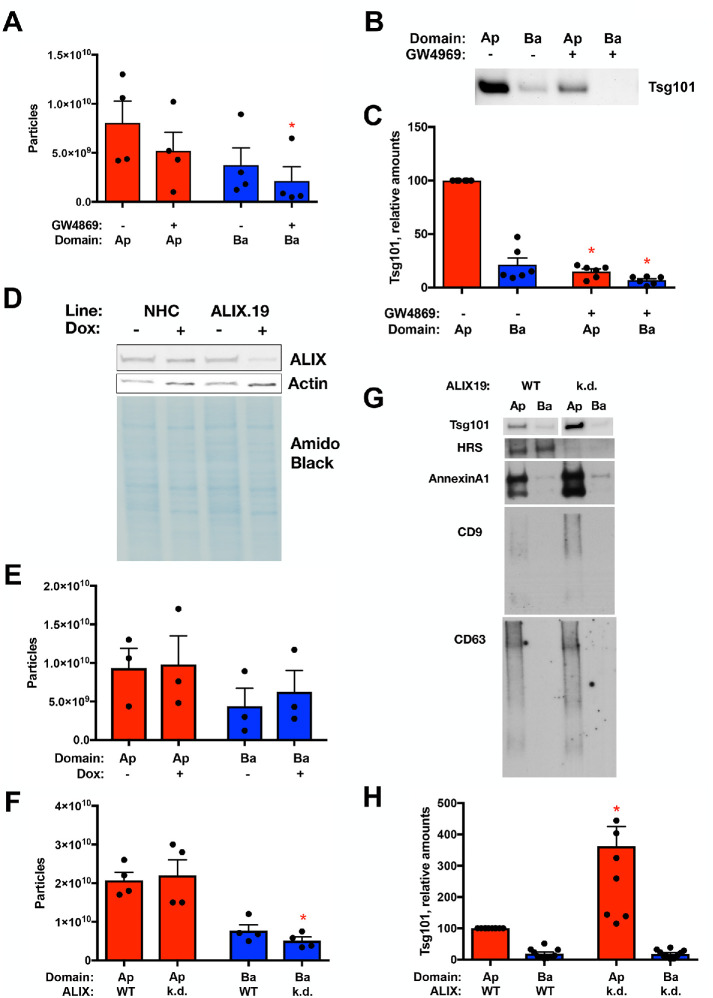
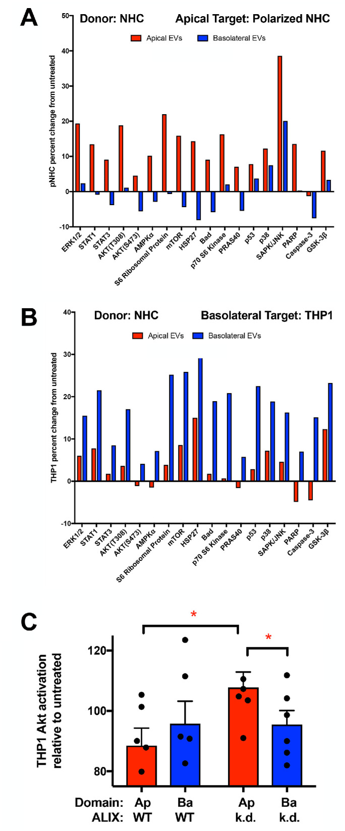
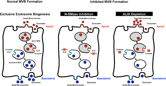
Intercellular communication is critical for organismal homeostasis, and defects can contribute to human disease states. Polarized epithelial cells execute distinct signaling agendas via apical and basolateral surfaces to communicate with different cell types. Small extracellular vesicles (sEVs), including exosomes and small microvesicles, represent an understudied form of intercellular communication in polarized cells. Human cholangiocytes, epithelial cells lining bile ducts, were cultured as polarized epithelia in a Transwell system as a model with which to study polarized sEV communication. Characterization of isolated apically and basolaterally released EVs revealed enrichment in sEVs. However, differences in apical and basolateral sEV composition and numbers were observed. Genetic or pharmacological perturbation of cellular machinery involved in the biogenesis of intralumenal vesicles at endosomes (the source of exosomes) revealed general and domain-specific effects on sEV biogenesis/release. Additionally, analyses of signaling revealed distinct profiles of activation depending on sEV population, target cell, and the function of the endosomal sorting complex required for transport (ESCRT)-associated factor ALG-2-interacting protein X (ALIX) within the donor cells. These results support the conclusion that polarized cholangiocytes release distinct sEV pools to mediate communication via their apical and basolateral domains and suggest that defective ESCRT function may contribute to disease states through altered sEV signaling.


Figures

FIGURE 1:
FIGURE 1:
Validation of polarized cholangiocyte Transwell culture system. (A) Transepithelial electrical resistance (TEER) was assessed to evaluate the formation of confluent NHC monolayers and the restricted current flow across the monolayer. Resistance is expressed as ohms•cm2. (B) Immunofluorescence staining of NHC Transwell cultures for Zonula occludens-1 (ZO1) indicated the formation of tight junctions between NHC cells grown in the Transwell culture system.
FIGURE 2:
FIGURE 2:
Polarized NHC release sEV particles from both apical and basolateral domains. (A) Western blotting equal volumes of apical and basolateral flotation fractions near the 10–20% interface for ESCRT components ALIX and Tsg101 and tetraspanin CD63 indicated peak reactivity in the 1.098 g/ml density fraction. (B) Size distribution resulting from NTA of the small EV fraction isolated from apical and basolateral conditioned media. Apical and basolateral EVs exhibit similar size distributions, and these profiles are consistent with exosome and small microvesicle particle enrichment. (C) Immunoelectron microscopy was performed with apical and basolateral EVs and antibody against the sEV marker CD63. Particles of sizes consistent with sEV and reactive with CD63 were observed in the EV fraction isolated from apical and basolateral conditioned media. (D) Western blotting equal volumes of apical and basolateral small EV fractions was performed with antibodies that detect markers of exosomes (Tsg101), microvesicles (Arf6), or Golgi (GM130). Whole cell extract (WC) was loaded as a positive control for immunodetection in addition to equal volumes of the apical (Ap) and basolateral (Ba) EV fractions. (E) Micrographs of NHC cell lines overexpressing Cherry-Rab27A wild type or dominant negative (DN, T21N), indicating expression in the majority of cells. Scale bar indicates 100 µm. (F) Western blots of NHC cell lines overexpressing Cherry-Rab27A wild type or dominant negative (DN, T21N), indicating enhanced expression. Endogenous Rab27A (darker exposure) and actin are indicated as loading controls. (G, H) Analysis of particles isolated from apical and basolateral conditioned media from NHC cell lines overexpressing Cherry-Rab27A wild type or dominant negative (DN, T21N). NTA (G) indicates reductions in apical (p value <0.05, paired t test) and basolateral (p value <0.05, unpaired t test) sEV numbers with Rab27A(DN) expression. Western blotting (H) indicates Rab27A(DN) reductions in ESCRT components ALIX and HRS and tetraspanin CD63 while the microvesicle marker AnnexinA1 was not impacted. The Golgi marker GM130 is presented as a control for contamination of the EV isolation.
FIGURE 3:
FIGURE 3:
Apical and basolateral small EV fractions exhibit differences in number and contents. (A) NTA total counts of small EV fraction particles isolated from the apical and basolateral conditioned media revealing that a greater number of particles within the apical chamber of polarized NHCs was apparent (n = 24, p value <0.05). (B) Total RNA concentration was assessed following isolation of RNA from equal numbers of apical and basolateral small EVs. Apical EV RNA content was elevated relative to basolateral EV RNA content (n = 10, p value <0.0001). (C–E) Cholesterol level was assessed and normalized to EV number (C), protein level (D), or phospholipid level (E). Apical EVs exhibited increased cholesterol per EV (n = 10, p value <0.02) and increased cholesterol:protein (n = 6, p value <0.03) and cholesterol:phospholipid (n = 5, p value <0.04) ratios. (F) Protein concentration was assessed and normalized to EV number (n = 27). (G) Extracts generated from equal numbers of apical and basolateral EVs were resolved by SDS–PAGE and silver stained. A number of species enriched in the apical (red) or basolateral (blue) EV samples are indicated for the purpose of illustration.
FIGURE 4:
FIGURE 4:
Apical and basolateral small EV miRNA contents differ. (A) miRNA profiling of the small EV fractions was performed using the BioWorks FireFly system, and the distribution of miRNA species between the apical (red) and basolateral (blue) EVs is presented. Asterisks indicate mRNA species further examined by QPCR in B. (B) Distributions of three miRNAs exhibiting differential enrichment by profiling were assessed by QPCR. miR34A exhibited apical enrichment (n = 3, p value <0.0001), miR486 exhibited weak apical enrichment (n = 3, p value <0.01), and miR223 exhibited basolateral enrichment (n = 3, p value <0.02).
FIGURE 5:
FIGURE 5:
Inhibition of N-SMase1 and depletion of ALIX exhibit different effects on polarized sEV release. (A–C) sEV release following treatment of polarized NHC with the N-SMase1 inhibitor GW4869 (10 µM) was assessed by NTA (A) and Western blotting for the ESCRT component Tsg101 loading equal volumes of the sEV fractions. (B, C) GW4869 treatment reduced basolateral EV particle numbers (n = 4, p value <0.05) and reduced Tsg101 levels in both the apical and basolateral EV fractions (n = 6, p value <0.0005 and <0.05, respectively). (D–H) sEV release following depletion of ALIX from polarized NHC. (D) Doxycycline-induced depletion in the ALIX.19 cell line was assessed by Western blotting for ALIX after 3 d treatment of polarized cells. Actin and amido black staining are presented as loading controls. (E) NTA of sEV release following doxycycline treatment of the parental NHC cell line, indicating similar release independent of treatment. (F–H) sEV release following depletion of ALIX from polarized NHC via doxycycline treatment was assessed by NTA (F) and Western blotting for the ESCRT components Tsg101 and HRS, the microvesicle marker AnnexinA1, and tetraspanins CD63 and CD9 loading equal volumes of the sEV fractions. (G, H). ALIX depletion reduced basolateral EV particle numbers (n = 4, p value <0.05) and increased Tsg101 levels in the apical EV fraction (n = 7, p value <0.005), correlating with increases in CD9 and AnnexinA1 in the apical sEV fraction.
FIGURE 6:
FIGURE 6:
Apical and basolateral EVs induce different signaling in apical and basolateral target cells. (A, B) Equal numbers of NHC-derived apical and basolateral sEVs were used to treat (A) polarized NHCs via the apical surface (apical target cell model) or (B) the human monocyte THP1 cell line (basolateral target cell model). Lysates were generated at 24 h, and signaling was assessed using the PathScan Intracellular Signaling Array and normalized to untreated samples. Apical sEVs induced greater activation in this apical target cell model (p value <0.0001), and basolateral sEVs induced greater activation in this basolateral target cell model (p value <0.01). (C) THP1 cells were treated with equal volumes of sEV fractions derived from NHC or ALIX-depleted ALIX.19 cells, lysates were generated at 24 h, and signaling was assessed by Western blotting for Akt activation and normalized to untreated control sample. Apical sEVs from ALIX-depleted cells induced greater Akt activation in this basolateral target cell model than either NHC-derived apical sEVs (n = 5, p value <0.05) or basolateral sEVs derived from ALIX-depleted cells. (n = 5, p value = 0.05).
FIGURE 7:
FIGURE 7:
Exclusive MVE biogenesis model. (Left) The sorting of exosome cargoes into MVEs occurs on distinct apical and basolateral endosomes, which then fuse with their respective plasma membrane domains to release the apical and basolateral exosomes. These exosomes along with small microvesicles shed from the plasma membrane comprise the sEV population. (Middle) Inhibition of the ceramide pathway via GW4869 treatment perturbed both the apical and basolateral sEV populations. (Right) In contrast, ALIX depletion reduced the numbers of basolateral sEVs released and altered the apical sEVs with respect to their contents and signaling capacity through reducing exosome secretion but enhancing small microvesicle shedding.

Similar articles

Cited by

References

    1. Al Suraih MS, Trussoni CE, Splinter PL, LaRusso NF, O’Hara SP (2020). Senescent cholangiocytes release extracellular vesicles that alter target cell phenotype via the epidermal growth factor receptor. Liver Int, DOI:10.1111/liv.14569. - DOI - PMC - PubMed
    1. Arbelaiz A, Azkargorta M, Krawczyk M, Santos-Laso A, Lapitz A, Perugorria MJ, Erice O, Gonzalez E, Jimenez-Aguero R, Lacasta A, et al (2017). Serum extracellular vesicles contain protein biomarkers for primary sclerosing cholangitis and cholangiocarcinoma. Hepatology , 1125–1143. - PubMed
    1. Bache KG, Slagsvold T, Cabezas A, Rosendal KR, Raiborg C, Stenmark H (2004a). The growth-regulatory protein HCRP1/hVps37A is a subunit of mammalian ESCRT-I and mediates receptor down-regulation. Mol Biol Cell , 4337–4346. - PMC - PubMed
    1. Bache KG, Slagsvold T, Stenmark H (2004b). Defective downregulation of receptor tyrosine kinases in cancer. EMBO J , 2707–2712. - PMC - PubMed
    1. Baietti MF, Zhang Z, Mortier E, Melchior A, Degeest G, Geeraerts A, Ivarsson Y, Depoortere F, Coomans C, Vermeiren E, et al (2012). Syndecan-syntenin-ALIX regulates the biogenesis of exosomes. Nat Cell Biol , 677–685. - PubMed

Publication types

LinkOut - more resources