Non-invasive prenatal testing (NIPT) by low coverage genomic sequencing: Detection limits of screened chromosomal microdeletions
- PMID: 32845907
- PMCID: PMC7449492
- DOI: 10.1371/journal.pone.0238245
Non-invasive prenatal testing (NIPT) by low coverage genomic sequencing: Detection limits of screened chromosomal microdeletions
Abstract
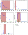
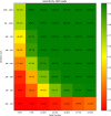
To study the detection limits of chromosomal microaberrations in non-invasive prenatal testing with aim for five target microdeletion syndromes, including DiGeorge, Prader-Willi/Angelman, 1p36, Cri-Du-Chat, and Wolf-Hirschhorn syndromes. We used known cases of pathogenic deletions from ISCA database to specifically define regions critical for the target syndromes. Our approach to detect microdeletions, from whole genome sequencing data, is based on sample normalization and read counting for individual bins. We performed both an in-silico study using artificially created data sets and a laboratory test on mixed DNA samples, with known microdeletions, to assess the sensitivity of prediction for varying fetal fractions, deletion lengths, and sequencing read counts. The in-silico study showed sensitivity of 79.3% for 10% fetal fraction with 20M read count, which further increased to 98.4% if we searched only for deletions longer than 3Mb. The test on laboratory-prepared mixed samples was in agreement with in-silico results, while we were able to correctly detect 24 out of 29 control samples. Our results suggest that it is possible to incorporate microaberration detection into basic NIPT as part of the offered screening/diagnostics procedure, however, accuracy and reliability depends on several specific factors.
Conflict of interest statement
I have read the journal's policy and the authors of this manuscript have the following competing interests: We declare potential competing financial interest in the form of employee contracts (see affiliations for each author) with Geneton Ltd. and TrisomyTest Ltd.. Geneton Ltd. participated in the development of a commercial NIPT test in Slovakia, however is not a provider of this commercial test, but still continues to do basic and applied research in the field of NIPT. On the other hand, TrisomyTest Ltd. is the commercial providers of NIPT testing in Slovakia, their participation in the study was, however, limited to the routine NIPT testing that generated the genomic results reused in our study. Related to this work, there are no patents, products in development or marketed products to declare. This does not alter our adherence to PLOS ONE policies on sharing data and materials, however, there are some restrictions in sharing of our data publicly. More details on these can be found in the Data Availability Statement.
Figures


Similar articles
-
BinDel: Detecting Clinically Relevant Fetal Genomic Microdeletions Using Low-Coverage Whole-Genome Sequencing-Based NIPT.Prenat Diagn. 2025 Mar;45(3):352-361. doi: 10.1002/pd.6758. Epub 2025 Feb 7. Prenat Diagn. 2025. PMID: 39921343 Free PMC article.
-
Positive predictive value estimates for cell-free noninvasive prenatal screening from data of a large referral genetic diagnostic laboratory.Am J Obstet Gynecol. 2017 Dec;217(6):691.e1-691.e6. doi: 10.1016/j.ajog.2017.10.005. Epub 2017 Oct 13. Am J Obstet Gynecol. 2017. PMID: 29032050
-
Noninvasive prenatal testing for microdeletion syndromes and expanded trisomies: proceed with caution.Obstet Gynecol. 2014 May;123(5):1097-1099. doi: 10.1097/AOG.0000000000000237. Obstet Gynecol. 2014. PMID: 24785862
-
Non-invasive prenatal testing for fetal chromosomal abnormalities by low-coverage whole-genome sequencing of maternal plasma DNA: review of 1982 consecutive cases in a single center.Ultrasound Obstet Gynecol. 2014 Mar;43(3):254-64. doi: 10.1002/uog.13277. Epub 2014 Feb 10. Ultrasound Obstet Gynecol. 2014. PMID: 24339153 Review.
-
Common genetic and epigenetic syndromes.Pediatr Clin North Am. 2015 Apr;62(2):411-26. doi: 10.1016/j.pcl.2014.11.005. Epub 2015 Jan 22. Pediatr Clin North Am. 2015. PMID: 25836705 Review.
Cited by
-
Evaluation of the clinical utility of extended non-invasive prenatal testing in the detection of chromosomal aneuploidy and microdeletion/microduplication.Eur J Med Res. 2023 Aug 30;28(1):304. doi: 10.1186/s40001-023-01285-2. Eur J Med Res. 2023. PMID: 37644576 Free PMC article.
-
Technical and Methodological Aspects of Cell-Free Nucleic Acids Analyzes.Int J Mol Sci. 2020 Nov 16;21(22):8634. doi: 10.3390/ijms21228634. Int J Mol Sci. 2020. PMID: 33207777 Free PMC article. Review.
-
Prenatal genetic diagnosis: Fetal therapy as a possible solution to a positive test.Acta Biomed. 2020 Nov 9;91(13-S):e2020021. doi: 10.23750/abm.v91i13-S.10534. Acta Biomed. 2020. PMID: 33170180 Free PMC article.
-
Prenatal sonographic findings in confirmed cases of Wolf-Hirschhorn syndrome.BMC Pregnancy Childbirth. 2022 Apr 15;22(1):327. doi: 10.1186/s12884-022-04665-4. BMC Pregnancy Childbirth. 2022. PMID: 35428251 Free PMC article.
-
BinDel: Detecting Clinically Relevant Fetal Genomic Microdeletions Using Low-Coverage Whole-Genome Sequencing-Based NIPT.Prenat Diagn. 2025 Mar;45(3):352-361. doi: 10.1002/pd.6758. Epub 2025 Feb 7. Prenat Diagn. 2025. PMID: 39921343 Free PMC article.
References
-
- Taylor-Phillips S, Freeman K, Geppert J, Agbebiyi A, Uthman OA, Madan J, et al. Accuracy of non-invasive prenatal testing using cell-free DNA for detection of Down, Edwards and Patau syndromes: a systematic review and meta-analysis. BMJ Open. 2016;6: e010002 10.1136/bmjopen-2015-010002 - DOI - PMC - PubMed
Publication types
MeSH terms
Substances
Supplementary concepts
LinkOut - more resources
Full Text Sources
Other Literature Sources
Research Materials

