. 2020 Aug 24;21(17):6088.
doi: 10.3390/ijms21176088.

Sephin1 Protects Neurons against Excitotoxicity Independently of the Integrated Stress Response

Affiliations

Sephin1 Protects Neurons against Excitotoxicity Independently of the Integrated Stress Response

Asier Ruiz et al. Int J Mol Sci. .

Abstract

Sephin1 is a derivative of guanabenz that inhibits the dephosphorylation of the eukaryotic initiation factor 2 alpha (eIF2α) and therefore may enhance the integrated stress response (ISR), an adaptive mechanism against different cellular stresses, such as accumulation of misfolded proteins. Unlike guanabenz, Sephin1 provides neuroprotection without adverse effects on the α2-adrenergic system and therefore it is considered a promising pharmacological therapeutic tool. Here, we have studied the effects of Sephin1 on N-methyl-D-aspartic acid (NMDA) receptor signaling which may modulate the ISR and contribute to excitotoxic neuronal loss in several neurodegenerative conditions. Time-course analysis of peIF2α levels after NMDA receptor overactivation showed a delayed dephosphorylation that occurred in the absence of activating transcription factor 4 (ATF4) and therefore independently of the ISR, in contrast to that observed during endoplasmic reticulum (ER) stress induced by tunicamycin and thapsigargin. Similar to guanabenz, Sephin1 completely blocked NMDA-induced neuronal death and was ineffective against AMPA-induced excitotoxicity, whereas it did not protect from experimental ER stress. Interestingly, both guanabenz and Sephin1 partially but significantly reduced NMDA-induced cytosolic Ca2+ increase, leading to a complete inhibition of subsequent calpain activation. We conclude that Sephin1 and guanabenz share common strong anti-excitotoxic properties with therapeutic potential unrelated to the ISR.

Keywords: NMDA; Sephin1; calcium; calpain; excitotoxicity; guanabenz; integrated stress response.


Conflict of interest statement

The authors declare no conflict of interest.

Figures

Figure 1
Figure 1
NMDAR activation induces activating transcription factor 4 (ATF4)-independent phosphorylation of eIF2α at Ser51 (peIF2α) dephosphorylation. (A) Cells were stimulated with NMDA (30 µM, 30 min) or thapsigargin (1 µM, always present) and harvested at indicated time points for immunoblotting of peIF2α, total eukaryotic initiation factor 2 (eIF2α), ATF4 and β-actin. (B) Quantification of peIF2α levels at indicated time points after NMDA treatment. Data represent normalized means ± SEM of three independent experiments. * p < 0.05; ** p < 0.01 compared with untreated cells (control), one-way ANOVA followed by Bonferroni´s post hoc test.
Figure 2
Figure 2
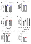
Sephin1 and guanabenz strongly attenuate NMDA-induced (but not AMPA/CTZ-induced) excitotoxicity. (A) Neurons were stimulated with NMDA (30 and 100 µM, 30 min) in the presence or absence of guanabenz (5 µM, present before, during, and after NMDA addition). * p < 0.05, *** p < 0.001 compared with control (NMDA alone), one-way ANOVA followed by Bonferroni´s post hoc test. (B) Neurons were treated with AMPA (25 µM, 30 min) and cyclothiazide (CTZ, 100 µM) in the presence or absence of guanabenz (5 µM, present before, during, and after AMPA/CTZ addition) and paired Student’s t-test. (C) Neurons were stimulated with NMDA (30 and 100 µM, 30 min) in the presence or absence of Sephin1 (5 µM, present before, during, and after NMDA addition). ** p < 0.01, **** p < 0.0001 compared with control (NMDA alone), one-way ANOVA followed by Bonferroni´s post hoc test. (D) Neurons were stimulated with 100 µM of NMDA for 30 min in the presence or absence of increasing concentrations of Sephin1 (1, 5, and 50 µM, added 1 h before NMDA addition and washed after NMDA). * p < 0.05 compared with control (NMDA alone), one-way ANOVA followed by Bonferroni´s post hoc test. (E) Neurons were stimulated with 100 µM of NMDA for 30 min in the presence or absence of Sephin1 (5 µM, added after NMDA wash). * p < 0.05 compared with control (NMDA alone), paired Student’s t-test. (F) Cells were treated with 25 µM of AMPA plus CTZ in the presence or absence of Sephin1 (5 µM, added 1 h before AMPA/CTZ addition), paired Student’s t-test. In all cases, cell viability was assessed 24 h later by the quantification of vital dye calcein-AM fluorescence. Data represent means ± SEM of normalized calcein fluorescence values.
Figure 3
Figure 3
Guanabenz but not Sephin1 reduces ER stress-induced neuronal death. Primary cultured neurons were exposed to tunicamycin and thapsigargin (1 µM both, 24 h) in the absence or presence of (A) guanabenz (5 µM, 24 h) and (B) Sephin1 (5 µM, 24 h). The number of viable cells was assessed by fluorescent vital dye calcein-AM 24 h later. Histograms represent means ± SEM of calcein fluorescence values normalized to controls. * p < 0.05 compared with cells treated with tunicamycin or thapsigargin alone, paired Student’s t-test.
Figure 4
Figure 4
Sephin1 and guanabenz reduce NMDA-induced cytosolic Ca2+ load. (A) Fluo-4-loaded neurons were exposed to NMDA (100 µM) in the absence or presence of guanabenz (5 µM) and cytosolic Ca2+ load quantified. *** p < 0.001 compared to control cells (NMDA alone), unpaired Student’s t-test. (B) Neurons were treated in the absence or presence of Sephin1 (1, 5 and 50 µM) and cytosolic Ca2+ load quantified. * p < 0.05, **** p < 0.0001 compared with control (NMDA alone), one-way ANOVA followed by Bonferroni´s post hoc test. Traces show normalized means ± SEM of several fields of view (n) from no less than three independent cultures. Statistical significance was calculated from normalized average ± SEM of the area under the curve.
Figure 5
Figure 5
Sephin1 and guanabenz inhibit NMDA-induced calpain activation. (A) Neurons were exposed to NMDA (100 µM, 30 min) in the absence or presence of Sephin1 or guanabenz (5 µM) and harvested 4 h later for the detection of αII-spectrin breakdown products (SBDP) and β-actin by Western blot. *** p < 0.001 compared with cells treated with NMDA alone (100%), paired Student’s t-test. (B) Neurons were treated with NMDA (100 µM, 30 min) in the absence or presence of increasing concentrations of Sephin1 (1, 5, 50 µM) and harvested as above. ** p < 0.05, **** p < 0.0001 compared with control (NMDA alone), one-way ANOVA followed by Bonferroni´s post hoc test. In all cases, for the quantification of calpain activity, SBDP150/145 signal was measured and normalized to β-actin values.

Similar articles

Cited by

References

    1. Luh L.M., Bertolotti A. Potential benefit of manipulating protein quality control systems in neurodegenerative diseases. Curr. Opin. Neurobiol. 2020;61:125–132. doi: 10.1016/j.conb.2020.02.009. - DOI - PubMed
    1. Pakos-Zebrucka K., Koryga I., Mnich K., Ljujic M., Samali A., Gorman A.M. The integrated stress response. EMBO Rep. 2016;17:1374–1395. doi: 10.15252/embr.201642195. - DOI - PMC - PubMed
    1. Harding H.P., Novoa I., Zhang Y., Zeng H., Wek R., Schapira M., Ron D. Regulated translation initiation controls stress-induced gene expression in mammalian cells. Mol. Cell. 2000;6:1099–1108. doi: 10.1016/S1097-2765(00)00108-8. - DOI - PubMed
    1. Harding H.P., Zhang Y., Zeng H., Novoa I., Lu P.D., Calfon M., Sadri N., Yun C., Popko B., Paules R., et al. An integrated stress response regulates amino acid metabolism and resistance to oxidative stress. Mol. Cell. 2003;11:619–633. doi: 10.1016/S1097-2765(03)00105-9. - DOI - PubMed
    1. Ma Y., Hendershot L.M. Delineation of a negative feedback regulatory loop that controls protein translation during endoplasmic reticulum stress. J. Biol. Chem. 2003;278:34864–34873. doi: 10.1074/jbc.M301107200. - DOI - PubMed

MeSH terms

LinkOut - more resources