. 2020 Aug 26;21(1):106.
doi: 10.1186/s10194-020-01174-7.

Optogenetic stimulation of the motor cortex alleviates neuropathic pain in rats of infraorbital nerve injury with/without CGRP knock-down

Affiliations

Optogenetic stimulation of the motor cortex alleviates neuropathic pain in rats of infraorbital nerve injury with/without CGRP knock-down

Jaisan Islam et al. J Headache Pain. .

Abstract

Background: Previous studies have reported that electrical stimulation of the motor cortex is effective in reducing trigeminal neuropathic pain; however, the effects of optical motor cortex stimulation remain unclear.

Objective: The present study aimed to investigate whether optical stimulation of the primary motor cortex can modulate chronic neuropathic pain in rats with infraorbital nerve constriction injury.

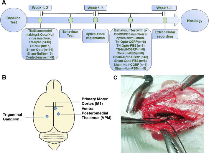
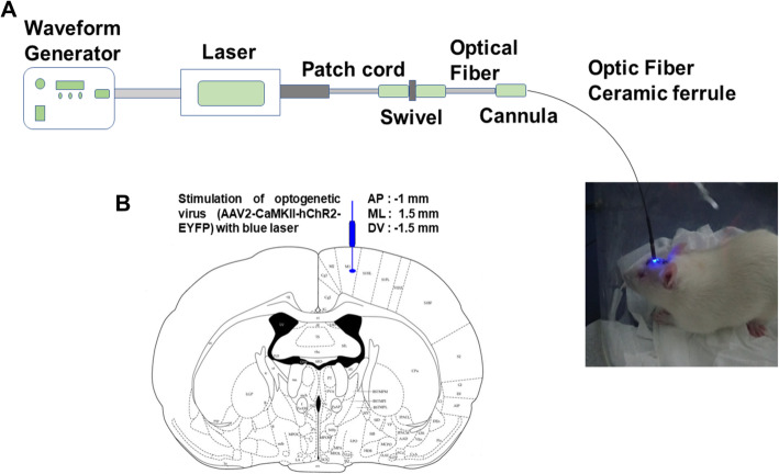
Methods: Animals were randomly divided into a trigeminal neuralgia group, a sham group, and a control group. Trigeminal neuropathic pain was generated via constriction of the infraorbital nerve and animals were treated via selective inhibition of calcitonin gene-related peptide in the trigeminal ganglion. We assessed alterations in behavioral responses in the pre-stimulation, stimulation, and post-stimulation conditions. In vivo extracellular recordings were obtained from the ventral posteromedial nucleus of the thalamus, and viral and α-CGRP expression were investigated in the primary motor cortex and trigeminal ganglion, respectively.

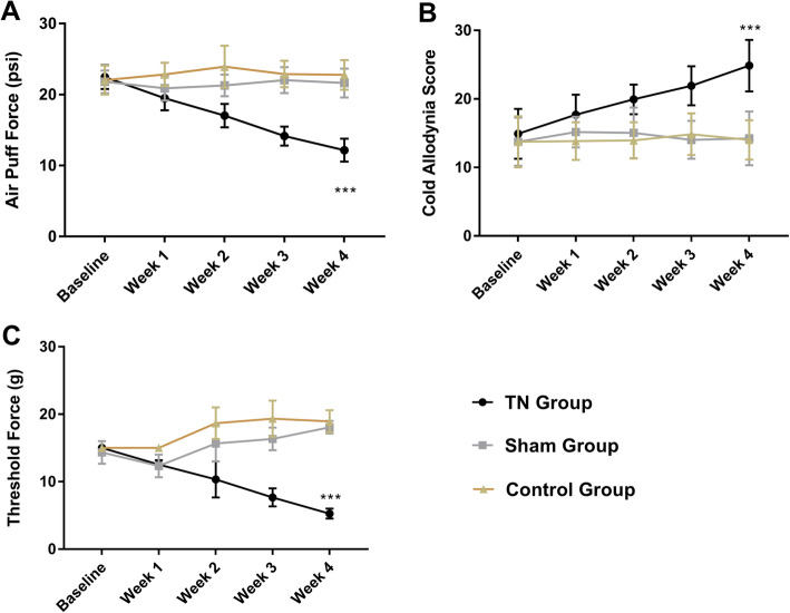
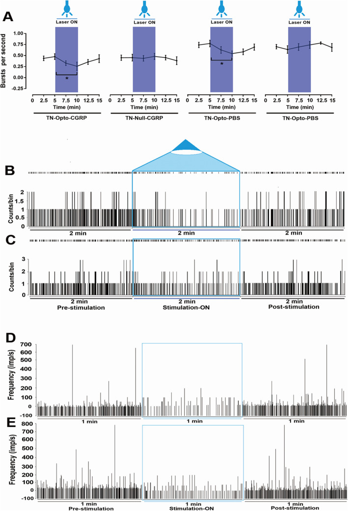
Results: We found that optogenetic stimulation significantly improved pain behaviors in the trigeminal neuralgia animals and it provided more significant improvement with inhibited α-CGRP state than active α-CGRP state. Electrophysiological recordings revealed decreases in abnormal thalamic firing during the stimulation-on condition.

Conclusion: Our findings suggest that optical motor cortex stimulation can alleviate pain behaviors in a rat model of trigeminal neuropathic pain. Transmission of trigeminal pain signals can be modulated via knock-down of α-CGRP and optical motor cortex stimulation.

Keywords: Motor cortex; Neuropathic pain; Optogenetics; Thalamus; Trigeminal ganglion; α-CGRP.


Conflict of interest statement

The authors have no conflicts of interest to declare.

Figures

Fig. 1
Fig. 1
Experimental animal model and timeline. a Experimental timeline. b Schematic diagram of the experimental animal model and optogenetic virus injection site. C. Ligation of the ION to produce the TN model. ION: infraorbital nerve, TN: trigeminal neuralgia
Fig. 2
Fig. 2
Optogenetic stimulation procedure. a Complete setup of optic stimulation. b Optic fiber implantation. c Stimulation of primary motor cortex using a blue laser
Fig. 3
Fig. 3
Behavioral test results for the TN, sham, and control groups. a Air-puff test results for the ipsilateral trigeminal facial area. b Cold hyperalgesia results for the ipsilateral facial area following treatment with acetone drops. c Mechanical allodynia (von Frey test) results for the ipsilateral facial side. ***, p < 0.001, significant difference determined via an analysis of variance (ANOVA)
Fig. 4
Fig. 4
Changes in behavioral responses following blue laser stimulation (a-c) along with knocking down of α-CGRP (d). Results for the air-puff test (a), cold hyperalgesia test (b), and von Frey test (c) for all eight animal groups. Only the TN-Opto-CGRP (a) and TN-Opto-PBS groups (b) exhibited significant changes in behavioral scores during the stimulation ON condition. No significant changes in behavior were observed in other animal groups during the stimulation ON condition. d Animals with α-CGRP injection exhibited more improvement in behavioral tests than animals with PBS injection. a Air puff force differences, b Cold hyperalgesia score differences and (c) Mechanical allodynia score differences between TN-Opto-CGRP and TN-Opto-PBS group. *, p < 0.05; **, p < 0.01, significant difference determined via an analysis of variance (ANOVA)
Fig. 5
Fig. 5
Electrophysiological findings for VPM thalamic output due to optogenetic stimulation of the motor cortex. a Evoked firing rates in VPM neurons in TN animals versus sham and control animals (***, p < 0.001, significant difference determined using unpaired t-tests). b, c In vivo recordings of the VPM thalamus from TN-Opto animals. b Findings for the TN-Opto-CGRP group. c Findings for the TN-Opto-PBS group. Decrease in firing output (spikes/s) were observed during the stimulation ON period. d, e No changes in firing rates of VPM thalamus were observed in the TN-Null-CGRP and TN-Null-PBS groups, respectively (two-way analysis of variance (ANOVA) *, p < 0.05; **, p < 0.01
Fig. 6
Fig. 6
Burst firing rates in TN animals following optogenetic stimulation (a). Significant changes were observed in the TN-Opto-CGRP and TN-Opto-PBS groups. *, p < 0.05; **, analysis of variance (ANOVA). b, c Peri-event raster histogram of VPM neuron responses in the TN-Opto-CGRP and TN-Opto-PBS groups, respectively. VPM firing rates decreases during optical stimulation. Bin size = 50 ms. d, e Instantaneous firing frequency for VPM neurons in TN-Opto-CGRP and TN-Opto-PBS rats, respectively
Fig. 7
Fig. 7
Immunofluorescence results for viral expression in the primary motor cortex (a-f) and CGRP neuron expression in the trigeminal ganglion (g-l). a, b, c Optogenetic viral expression in the motor cortex of TN-Opto animals. d, e, f Null viral expression in the motor cortex of TN-Null animals. a, d EYFP, b, e DAPI, and c, f Merge. Scale bar = 200 μm. g-l PBS and α-CGRP-injected and DAPI-stained trigeminal ganglion cells. g-i Presence of anti-CGRP antibody binding due to active state of α-CGRP neuron (arrow) within the trigeminal ganglion of PBS injected animal (j-l). α-CGRP injected and DAPI-stained trigeminal ganglion cells showing absence of CGRP neurons due to inhibition of α-CGRP neurons within the trigeminal ganglion. g, j CGRP, h, k DAPI, and i, l Merge. Scale bar = 100 μm

References

    1. Henderson JM, Lad SP. Motor cortex stimulation and neuropathic facial pain. Neurosurg Focus. 2006;21(6):1–4. - PubMed
    1. Moon HC, Heo WI, Kim YJ, Lee D, Won SY, Kim HR, et al. Optical inactivation of the anterior cingulate cortex modulate descending pain pathway in a rat model of trigeminal neuropathic pain created via chronic constriction injury of the infraorbital nerve. J Pain Res. 2017;10:2355. - PMC - PubMed
    1. Saavedra LC, Mendonca M, Fregni F. Role of the primary motor cortex in the maintenance and treatment of pain in fibromyalgia. Med Hypotheses. 2014;83(3):332–336. - PubMed
    1. Fontaine D, Hamani C, Lozano A. Efficacy and safety of motor cortex stimulation for chronic neuropathic pain: critical review of the literature. J Neurosurg. 2009;110(2):251–256. - PubMed
    1. Delavallee M, Rooijakkers H, Koerts G, Raftopoulos C. Motor cortex stimulation in a three-year-old child with trigeminal neuropathic pain caused by a malignant glioma in the cerebellopontine angle: case report. Neurosurgery. 2011;69(2):E494–E4E6. - PubMed

Substances