. 2020 Aug 26;20(1):100.
doi: 10.1186/s12880-020-00498-9.

Myofibroma/myofibromatosis: MDCT and MR imaging findings in 24 patients with radiological-pathological correlation

Affiliations

Myofibroma/myofibromatosis: MDCT and MR imaging findings in 24 patients with radiological-pathological correlation

Kun-Ming Yi et al. BMC Med Imaging. .

Abstract

Background: The aim of this study was to characterize the radiological features of myofibroma on multidetector computed tomography (MDCT) and magnetic resonance imaging (MRI) and correlate the imaging findings with pathologic features.

Methods: The radiological findings of 24 patients with 29 myofibromas were retrospectively reviewed. All images were evaluated with emphasis on density, signal intensity, hypointense area, and enhancement, correlating these with pathologic findings.

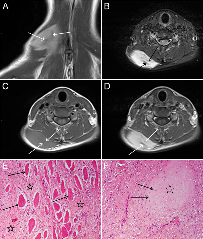
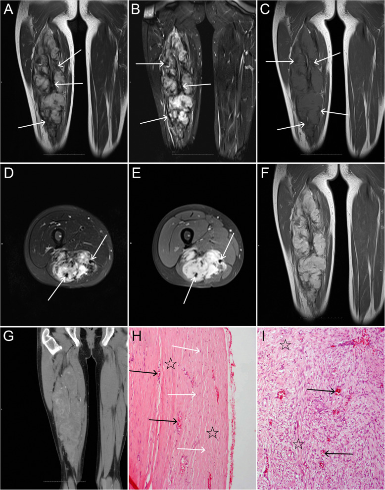
Results: On plain MDCT scan, 4(26.7%) tumors were homogeneous isodensity, 4(26.7%) tumors were heterogeneous hyperdensity, and 7(46.7%) tumors were heterogeneous hypodensity. On contrast-enhanced MDCT scan, all tumors (9/9) showed heterogeneous enhancement with moderate in 3(33.3%) and marked in 6(66.7%) tumors, and their enhancements were higher compared to adjacent skeletal muscle (P = 0.0001). On MRI, heterogeneous slight hyperintensity, homogeneous slight hyperintensity, and heterogeneous hypointensity on T1-weighted imaging (T1WI) were observed in 14(82.3%), 1(5.9%) and 2(11.8%) tumors, respectively. On T2-weighted imaging (T2WI) and fat-suppressed (FS) T2WI, all tumors demonstrated heterogeneous hyperintensity. All tumors showed heterogeneous marked enhancement on FS contrast-enhanced T1WI. On T1WI, T2WI, FS T2WI, and FS contrast-enhanced T1WI, irregular strip or/and patchy hypointensities were found in 16(94.1%), 12(100%), 17(100%) and 17(100%) tumors, respectively, and pseudocapsule was seen in 5(29.4%) tumors. The hypointensities and pseudocapsule on MRI were exactly corresponding to pathological interlacing collagen fibers and fibrosis. The age of the recurrent group was lower than that of the non-recurrent group (P = 0.001) and the tumors without pseudocapsule were more likely to recur than those with pseudocapsule (P = 0.034).

Conclusion: Myofibromas are characterized by heterogeneous density or signal intensity, with moderate or marked enhancement. The hypointensities and pseudocapsule on MRI may be helpful in diagnosis, and the absence of pseudocapsule and younger age may be risk factors for tumor recurrence.

Keywords: Magnetic resonance imaging; Multidetector computed tomography; Myofibroma; Myofibromatosis; Pathology.


Conflict of interest statement

The authors declare that they have no competing interests.

Figures

Fig. 1
Fig. 1
A young patient in his twenties with a myofibroma in the right body of the mandible. a and b: Axial unenhanced MDCT images show a well-defined round lesion on the lateral margin of the mandible, with periosteal reaction (white arrows) and resorption of the external cortical bone (black arrows). c and d: The lesion shows marked enhancement on axial arterial- and venous-phase contrast-enhanced MDCT images and greater enhancement is seen in the venous phase. e: Photomicrograph (HE, × 100) demonstrates a large number of interlacing bundles of spindle-shaped cells (☆) with tapered blunt-ended nuclei and eosinophilic cytoplasm; hemangiopericytoma-like pattern of the blood vessels (arrows) is focally seen. f: Inflammatory cells (☆) are also present in the lesion (HE, × 100)
Fig. 2
Fig. 2
A young patient in her twenties with a myofibroma in the right trapezius. a and b: Coronal T2WI and axial FS T2WI demonstrate a lobulated, heterogeneous hyperintense mass with irregular strip hypointensities (arrows). c: On axial T1WI, the lesion appears as heterogeneous slight hyperintensity with irregular hypointense areas (arrows). d: On contrast-enhanced axial FS T1WI, the lesion shows inhomogeneous marked enhancement with non-enhanced hypointense areas. e: Photomicrograph (HE, × 100) shows a large number of interlacing bundles of spindle-shaped cells (☆) between normal muscles (arrows). f: Local hyalinization (☆) with a few degenerative tumor cells (arrows) can be seen (HE, × 100)
Fig. 3
Fig. 3
A young teenage patient with a myofibroma in the right thigh. a and b: Coronal T2WI and FS T2WI reveal a huge lobulated, heterogeneous hyperintense mass with irregular patchy, strip hypointensities, and incomplete pseudocapsule (arrows). c: Coronal T1WI shows heterogeneous slight hyperintensity with irregular hypointense areas (arrows). d and e: Axial T2WI and contrast-enhanced FS T1WI reveal the lesion encircling tendon (arrows). e and f: On contrast-enhanced axial FS T1WI and coronal T1WI, the lesion shows inhomogeneous marked enhancement with non-enhanced hypointense areas. g: On contrast-enhanced MDCT image, the lesion shows heterogeneous enhancement. h: Photomicrograph (HE, × 100) shows a pseudocapsule consisting of a large number of collagen fibers (☆) with a small number of spindle cells (white arrows) and vascular proliferation (black arrows) in the lesion. i: A large amount of vascular proliferation (arrows) and irregular fibrosis (☆) are present in the center of the lesion (HE, × 100)

References

    1. Chung EB, Enzinger FM. Infantile myofibromatosis. Cancer. 1981;48:1807–1818. doi: 10.1002/1097-0142(19811015)48:8<1807::AID-CNCR2820480818>3.0.CO;2-G. - DOI - PubMed
    1. Matthews MR, Cockerell CJ. An historic perspective of infantile myofibromatosis. Adv Dermatol. 2006;22:279–305. doi: 10.1016/j.yadr.2006.09.003. - DOI - PubMed
    1. Mentzel T, Dei Tos AF, Sapi Z, Kutzner H. Myopericytoma of skin and soft tissues: Clinicopathologic and immunohistochemical study of 54 cases. Am J Surg Pathol. 2006;30:104–113. doi: 10.1097/01.pas.0000178091.54147.b1. - DOI - PubMed
    1. Fletcher CD, Hogendoorn PC, Mertens F, Bridge J. WHO classification of tumors of soft tissue and bone. 4. Lyon: IARC Press; 2013.
    1. Martignetti JA, Tian L, Li D, et al. Mutations in PDGFRB cause autosomal-dominant infantile myofibromatosis. Am J Hum Genet. 2013;92:1001–1007. doi: 10.1016/j.ajhg.2013.04.024. - DOI - PMC - PubMed

Publication types