Case Reports
. 2020 Aug 26;8(1):147.
doi: 10.1186/s40478-020-01024-2.

Neuronophagia and microglial nodules in a SARS-CoV-2 patient with cerebellar hemorrhage

Affiliations
Case Reports

Neuronophagia and microglial nodules in a SARS-CoV-2 patient with cerebellar hemorrhage

Osama Al-Dalahmah et al. Acta Neuropathol Commun. .

Abstract

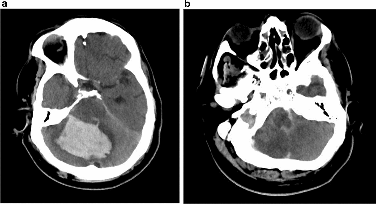
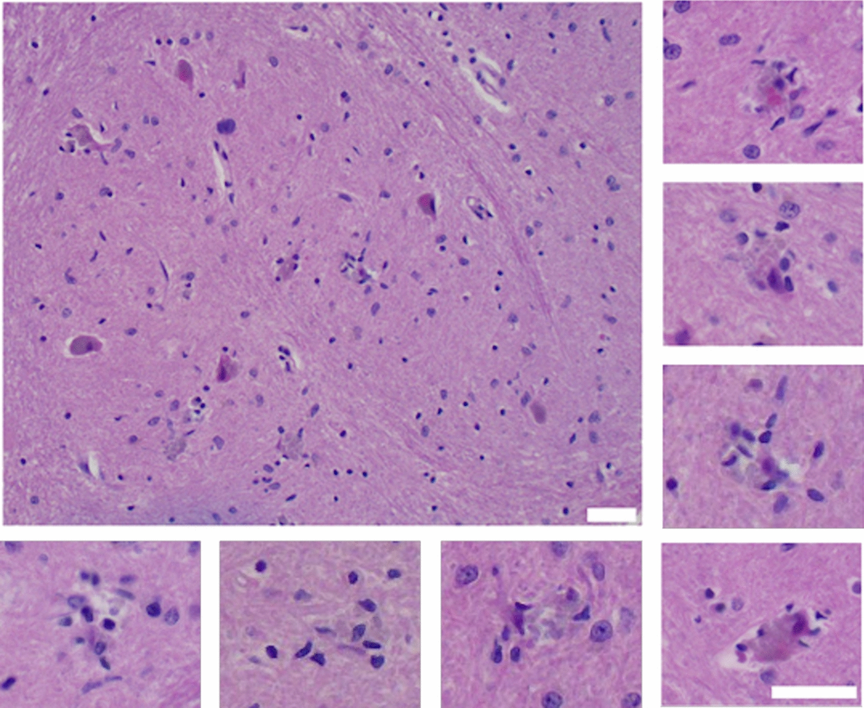
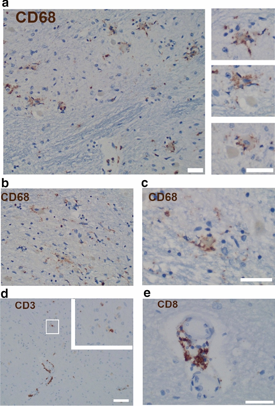
We document the neuropathologic findings of a 73-year old man who died from acute cerebellar hemorrhage in the context of relatively mild SARS-CoV2 infection. The patient developed sudden onset of headache, nausea, and vomiting, immediately followed by loss of consciousness on the day of admission. Emergency medical services found him severely hypoxemic at home, and the patient suffered a cardiac arrest during transport to the emergency department. The emergency team achieved return of spontaneous circulation after over 17 min of resuscitation. A chest radiograph revealed hazy bilateral opacities; and real-time-PCR for SARS-CoV-2 on the nasopharyngeal swab was positive. Computed tomography of the head showed a large right cerebellar hemorrhage, with tonsillar herniation and intraventricular hemorrhage. One day after presentation, he was transitioned to comfort care and died shortly after palliative extubation. Autopsy performed 3 h after death showed cerebellar hemorrhage and acute infarcts in the dorsal pons and medulla. Remarkably, there were microglial nodules and neuronophagia bilaterally in the inferior olives and multifocally in the cerebellar dentate nuclei. This constellation of findings has not been reported thus far in the context of SARS-CoV-2 infection.

Keywords: COVID-19; Microglial nodules; Neuronophagia; Neuropathology; SARS-CoV-2.


Conflict of interest statement

The author declares that they have no competing interest.

Figures

Fig. 1
Fig. 1
Axial non-contrasted head CT scans of the posterior fossa. The images show a large cerebellar hematoma with surrounding edema (a) as well as edema in the medulla (b)
Fig. 2
Fig. 2
Microglial nodules in the inferior olivary nuclei. Representative images of the inferior olivary nuclei showing microglial nodules and neuronophagia. The large panel (top left) shows an overall view of the left inferior olivary nucleus, with microglial nodules and neuronal loss. Several olivary neurons appear hyper-eosinophilic. The insets show higher power images of individual examples of microglial nodules with varying degrees of neuronal damage. Scale bars measure 100 µm
Fig. 3
Fig. 3
Neuronophagia in the inferior olivary and the dentate nuclei. a Neuronophagia highlighted by a CD68 immunohistochemical stain. Note the clusters of activated microglia surrounding shrunken and apparently normal inferior olivary neurons. Higher magnification images are shown on the right. b, c Immunohistochemical stain for CD68 showing microglial activation and neuronophagia in the cerebellar dentate nucleus. d Immunohistochemical stain for CD3 highlighted perivascular lymphocytes around venules in the medulla and sparse parenchymal infiltrates. The inset shows an example of a T cell juxtaposed to an olivary neuron. e Immunohistochemical stain for CD8 in the cerebellar white matter shows a subset of perivascular lymphocytes were CD8 positive. No CD8 lymphocytes were seen in the parenchyma. Scale bars measure 100 µm

References

    1. Adachi T, Chong JM, Nakajima N, Sano M, Yamazaki J, Miyamoto I, Nishioka H, Akita H, Sato Y, Kataoka M, et al. Clinicopathologic and immunohistochemical findings from autopsy of patient with COVID-19, Japan. Emerg Infect Dis. 2020 doi: 10.3201/eid2609.201353. - DOI - PMC - PubMed
    1. Barton LM, Duval EJ, Stroberg E, Ghosh S, Mukhopadhyay S. COVID-19 autopsies, Oklahoma, USA. Am J Clin Pathol. 2020;153:725–733. doi: 10.1093/ajcp/aqaa062. - DOI - PMC - PubMed
    1. Beyrouti R, Adams ME, Benjamin L, Cohen H, Farmer SF, Goh YY, Humphries F, Jäger HR, Losseff NA, Perry RJ, et al. Characteristics of ischaemic stroke associated with COVID-19. J Neurol Neurosurg Psychiatry. 2020 doi: 10.1136/jnnp-2020-323586. - DOI - PMC - PubMed
    1. Brown GC, Neher JJ. Microglial phagocytosis of live neurons. Nat Rev Neurosci. 2014;15:209–216. doi: 10.1038/nrn3710. - DOI - PubMed
    1. Chen N, Zhou M, Dong X, Qu J, Gong F, Han Y, Qiu Y, Wang J, Liu Y, Wei Y, et al. Epidemiological and clinical characteristics of 99 cases of 2019 novel coronavirus pneumonia in Wuhan, China: a descriptive study. Lancet. 2020;395:507–513. doi: 10.1016/S0140-6736(20)30211-7. - DOI - PMC - PubMed

Publication types

MeSH terms