

Cardiac papillary fibroelastoma originating from the coumadin ridge and review of literature
- PMID: 32847879
- PMCID: PMC7451947
- DOI: 10.1136/bcr-2020-235361
Cardiac papillary fibroelastoma originating from the coumadin ridge and review of literature
Abstract
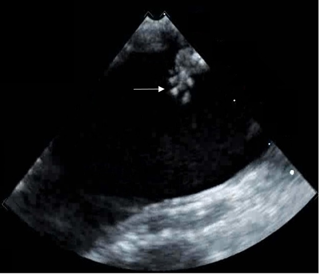
Papillary fibroelastomas represent the second most common benign cardiac tumour, secondary only to cardiac myxoma. A majority of patients are asymptomatic on presentation. The most common clinical manifestations include stroke, transient ischaemic attack, myocardial infarction and angina. Echocardiography remains the primary imaging modality for identification of these tumours. The majority of papillary fibroelastomas arise from the valves. Simple surgical excision is the mainstay of treatment, carrying an excellent prognosis. We present an unusual case of cardiac papillary fibroelastoma originating from the coumadin ridge (CR) in a 70-year-old woman. The patient exhibited increasing paroxysms of her atrial fibrillation and was pursuing a MAZE procedure. Preoperatively, a transesophageal echocardiogram revealed a 0.7×1 cm intracardiac mass that had echocardiographic appearance of a fibroelastoma. Surgical resection and MAZE procedures were performed. The gross specimen and histopathology findings were consistent with papillary fibroelastoma. This case reports the seventh documented case of fibroelastoma originating from the CR.
Keywords: cancer - see oncology; cardiothoracic surgery; interventional cardiology; surgical oncology.
© BMJ Publishing Group Limited 2020. No commercial re-use. See rights and permissions. Published by BMJ.
Conflict of interest statement
Competing interests: None declared.
Figures


References
Publication types
MeSH terms
LinkOut - more resources
Full Text Sources