. 2020 Aug 26;5(4):e00748-20.
doi: 10.1128/mSphere.00748-20.

Antibiotic Resistance in Vibrio cholerae: Mechanistic Insights from IncC Plasmid-Mediated Dissemination of a Novel Family of Genomic Islands Inserted at trmE

Affiliations

Antibiotic Resistance in Vibrio cholerae: Mechanistic Insights from IncC Plasmid-Mediated Dissemination of a Novel Family of Genomic Islands Inserted at trmE

Nicolas Rivard et al. mSphere. .

Abstract

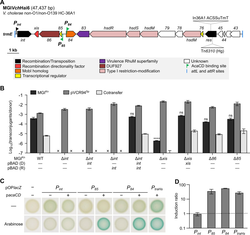
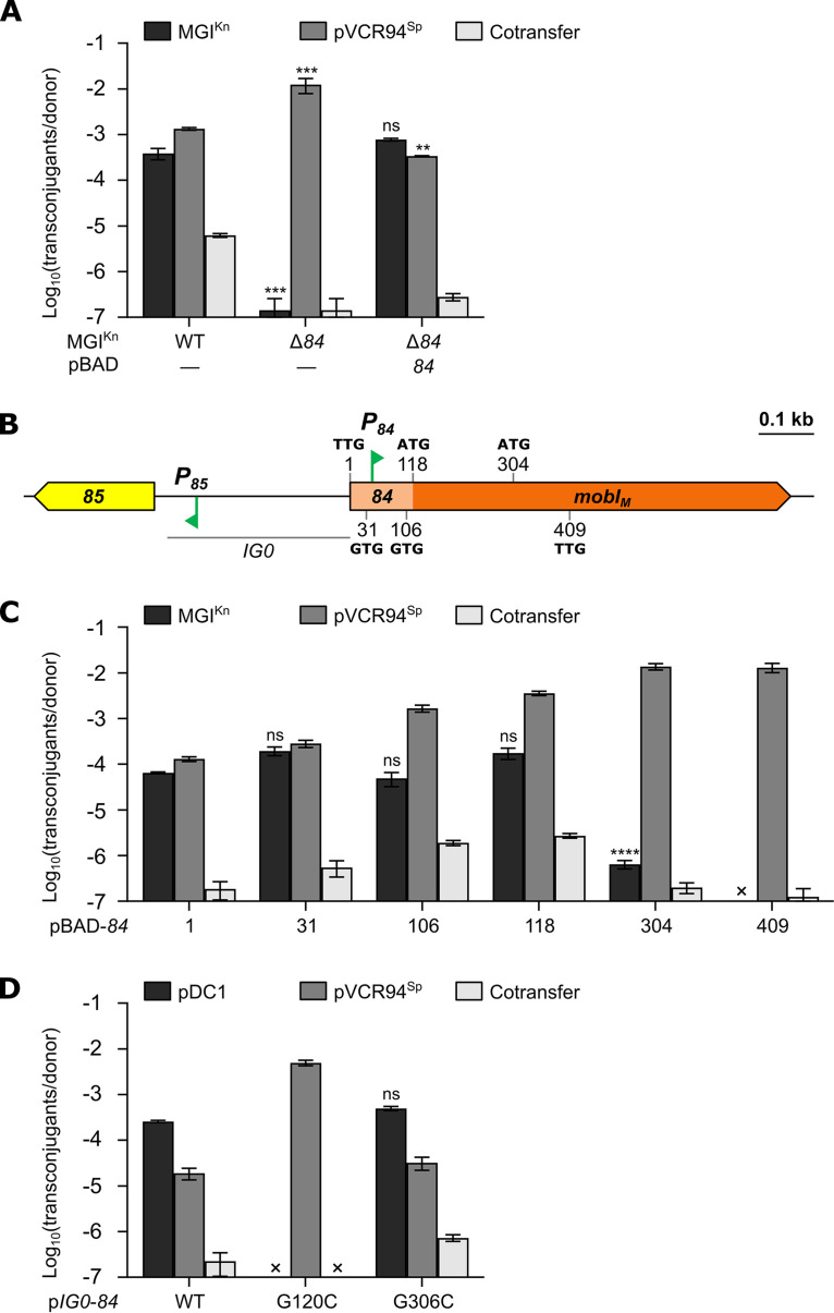
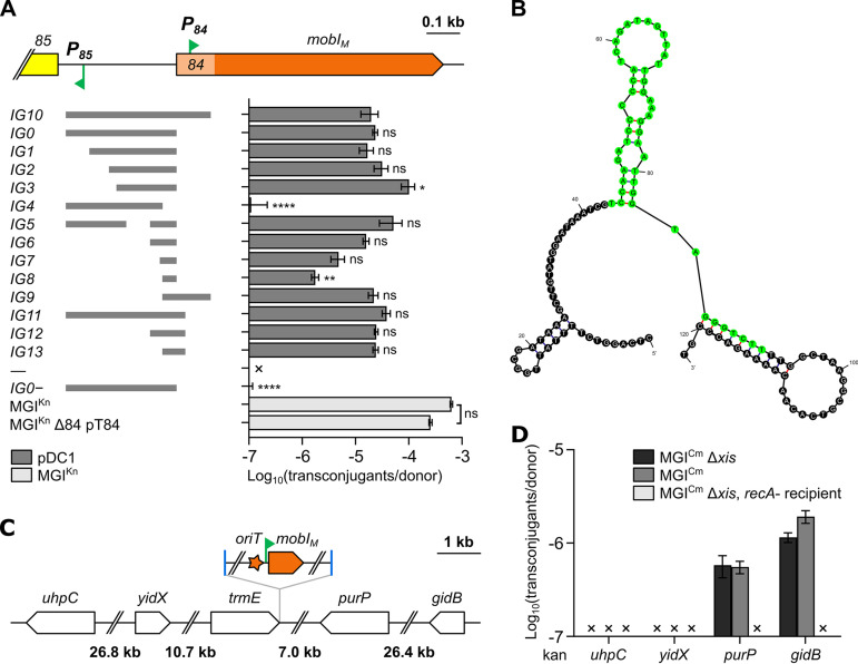
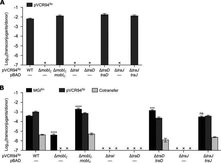
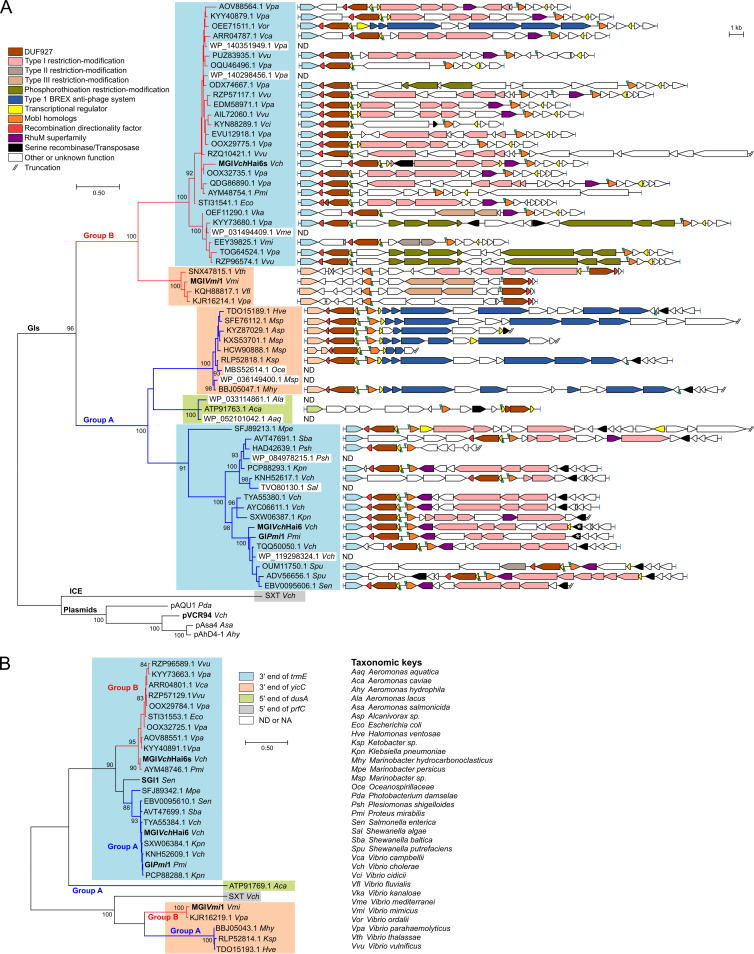
Cholera remains a formidable disease, and reports of multidrug-resistant strains of the causative agent Vibrio cholerae have become common during the last 3 decades. The pervasiveness of resistance determinants has largely been ascribed to mobile genetic elements, including SXT/R391 integrative conjugative elements, IncC plasmids, and genomic islands (GIs). Conjugative transfer of IncC plasmids is activated by the master activator AcaCD whose regulatory network extends to chromosomally integrated GIs. MGIVchHai6 is a multidrug resistance GI integrated at the 3' end of trmE (mnmE or thdF) in chromosome 1 of non-O1/non-O139 V. cholerae clinical isolates from the 2010 Haitian cholera outbreak. In the presence of an IncC plasmid expressing AcaCD, MGIVchHai6 excises from the chromosome and transfers at high frequency. Herein, the mechanism of mobilization of MGIVchHai6 GIs by IncC plasmids was dissected. Our results show that AcaCD drives expression of GI-borne genes, including xis and mobIM , involved in excision and mobilization. A 49-bp fragment upstream of mobIM was found to serve as the minimal origin of transfer (oriT) of MGIVchHai6. The direction of transfer initiated at oriT was determined using IncC plasmid-driven mobilization of chromosomal markers via MGIVchHai6. In addition, IncC plasmid-encoded factors, including the relaxase TraI, were found to be required for GI transfer. Finally, in silico exploration of Gammaproteobacteria genomes identified 47 novel related and potentially AcaCD-responsive GIs in 13 different genera. Despite sharing conserved features, these GIs integrate at trmE, yicC, or dusA and carry a diverse cargo of genes involved in phage resistance.IMPORTANCE The increasing association of the etiological agent of cholera, Vibrio cholerae serogroup O1 and O139, with multiple antibiotic resistance threatens to deprive health practitioners of this effective tool. Drug resistance in cholera results mainly from acquisition of mobile genetic elements. Genomic islands conferring multidrug resistance and mobilizable by IncC conjugative plasmids were reported to circulate in non-O1/non-O139 V. cholerae clinical strains isolated from the 2010 Haitian cholera outbreak. As these genomic islands can be transmitted to pandemic V. cholerae serogroups, their mechanism of transmission needed to be investigated. Our research revealed plasmid- and genomic island-encoded factors required for the resistance island excision, mobilization, and integration, as well as regulation of these functions. The discovery of related genomic islands carrying diverse phage resistance genes but lacking antibiotic resistance-conferring genes in a wide range of marine dwelling bacteria suggests that these elements are ancient and recently acquired drug resistance genes.

Keywords: IncC plasmids; T4CP; T4SS; Vibrio cholerae; antibiotic resistance; conjugation; genomic islands; horizontal gene transfer; mobilization; oriT; phage resistance; relaxase.


Figures

FIG 1
FIG 1
Role and regulation of int and 85-86-xis in the mobilization of MGIVchHai6. (A) Schematic genetic map of MGIVchHai6 drawn to scale. The left and right junctions (attL and attR) within the host chromosome are indicated by blue ticks at the extremities. ORFs with similar function are color coded as indicated in the figure. Green flags indicate the position and orientation of predicted AcaCD binding sites (13). The black flag indicates the position and orientation of the Pint promoter. The insertion sites of In36A1 integron and Tn6310 transposon are shown. The gene numbers correspond to the last digits of the respective locus tags in GenBank accession no. AXDR01000001. ACSSuTmT, resistance to ampicillin, chloramphenicol, spectinomycin/streptomycin, sulfamethoxazole, trimethoprim, and tetracycline; Hg, mercury resistance. (B) Mobilization assays of MGIKn or its Δint, Δxis, Δ86, or Δ85 mutants were carried out using E. coli GG56 (Nx) bearing pVCR94Sp as the donor strain, and CAG18439 (Tc) as the recipient strain. Complementation assays were performed in the donor (D) or recipient (R) strain by expressing the missing gene from PBAD on pBAD-int or pBAD-xis. An “×” indicates that the transfer frequency was below the detection limit (<10−7). Bars represent the means ± standard errors of the means (error bars) from three independent experiments. Statistical analyses were carried out on the logarithm of the values using a one-way analysis of variance (ANOVA), followed by Dunnett’s multiple-comparison test with the wild-type (WT) MGIKn as the control. Statistical significance is indicated as follows: ****, P < 0.0001; ns, not significant. (C) β-Galactosidase activities of Pint, P85, and P84 transcriptionally fused to lacZ. Colonies were grown on LB agar with or without arabinose to induce acaCD expression from pacaCD. (D) Induction levels of Pint, P85, and P84 in response to AcaCD. β-Galactosidase assays were carried out using the strains of panel C. Ratios between the enzymatic activities in Miller units for the arabinose-induced versus noninduced strains containing pacaCD are shown. The AcaCD-regulated promoter PtraHs of SGI1 served as a positive control, and cells devoid of pOPlacZ served as a negative control. The bars represent the means ± standard errors of the means (error bars) from two independent experiments.
FIG 2
FIG 2
Role of 84 in MGIVchHai6 mobilization. (A) Mobilization of MGIKn or its Δ84 mutant by pVCR94Sp. (B) Schematic representation of the 85-84 region of MGIVchHai6. Genes are color coded as indicated in Fig. 1A. The position and sequence of alternative start codons within 84 are indicated. (C) Complementation assays of the Δ84 mutation by alternative ORFs within 84. When indicated, donor strains contained the complementation plasmid pBAD-84 or one of its derivatives (Table 1). (D) Confirmation of ATG118 as the genuine start codon of mobIM. Conjugation assays were performed using E. coli GG56 (Nx) containing the specified elements as donor strains and either CAG18439 (Tc) (A and C) or VB112 (Rf) (D) as the recipient strain. The bars represent the means ± standard errors of the means from three independent experiments. Statistical analyses were carried out on the logarithm of the values using a one-way ANOVA followed by Dunnett’s multiple-comparison test with the WT MGIKn (A), pBAD-84 (C), or pIG0-84 (D) as the control. Statistical significance is indicated as follows: ****, P < 0.0001; ***, P < 0.001; **, P < 0.01; ns, not significant.
FIG 3
FIG 3
Localization of the oriT locus of MGIVchHai6. (A) On the left, fragments of the 85-mobI region that were cloned into the nonmobilizable vector pDC1 are represented by gray bars. The resulting transfer frequencies for the corresponding fragments are presented on the right side. Mobilization assays of pDC1 derivatives were performed using E. coli GG56 (Nx) bearing pVCR94Sp and pT84 as the donor and VB112 (Rf) as the recipient. “—” indicates an empty pDC1. “IG0−” indicates that the transfer of pIG0 was assessed in the absence of pT84. Statistical analyses were carried out on the logarithm of the values using a one-way ANOVA followed by Dunnett’s multiple-comparison test with pIG10 or MGIKn as the control. Statistical significance is indicated as follows: ****, P < 0.0001; **, P < 0.01; *, P < 0.05; ns, not significant. (B) Predicted folding of IG12, with IG8 highlighted in green. Folding of the upper strand (panel A) was predicted using the Mfold web server (74). (C) Schematic map of the chromosomal region surrounding trmE in E. coli K-12. The position and orientation of MGICm are indicated. (D) MGICm-mediated mobilization of chromosomal markers from E. coli JW3642, JW3692, JW3718, or JW5858 (Kn) bearing pVCR94Sp and MGICm or its Δxis mutant to CAG18439 or its ΔrecA mutant (Tc). In panels A and D, the bars represent the means ± standard errors of the means from three independent experiments. “×” indicates that the transfer frequency was below the detection limit (<10−7).
FIG 4
FIG 4
Mobilization of MGIVchHai6 relies on the T4CP and relaxase encoded by IncC plasmids. (A) Impact of mobIC, traI, traD, and traJ deletions on pVCR94Sp transfer. (B) Mobilization of MGIKn by the mobIC, traI, traD, and traJ deletion mutants of pVCR94Sp. Conjugation assays were performed with E. coli GG56 (Nx) containing the specified elements as donor strains and CAG18439 (Tc) as the recipient strain. The bars represent the means ± standard errors of the means from three independent experiments. “×” indicates that the transfer frequency was below the detection limit (<10−7). Statistical analyses were carried out on the logarithm of the values using a one-way ANOVA followed by Dunnett’s multiple-comparison test with WT MGIKn as the control. Statistical significance is indicated as follows: ****, P < 0.0001; ***, P < 0.001; ns, not significant.
FIG 5
FIG 5
Diversity of genomic islands encoding MobI homologs. (A and B) Maximum likelihood phylogenetic analysis of MobI (A) and Int (B) homologs. Trees with the highest likelihoods (−7,967.92 and −5,688.94 for MobI and Int, respectively) are shown. Bootstrap supports are indicated as percentages at the branching points only when >80%. Branch lengths represent the number of substitutions per site over 201 and 311 amino acid positions for MobI and Int, respectively. Only one representative per cluster of similar proteins (>95% identity threshold) is shown in each tree. In panel A, the schematic structure of the genomic island encoding the corresponding MobI protein is shown next to each node. ORFs with similar function are color coded as indicated in the panel. Each node and the integrase gene of the corresponding node are color coded based on the integration site of the genomic island (refer to panel B for color key [ND, not determined; NA, not available]). Vertical green arrowheads indicate position and orientation of AcaCD binding sites. attL and attR attachment sites flanking each GI are represented by blue bars. The asterisks in MGIVchHai6 and GIPmi1 indicate the insertion site of the complex resistance integrons. Additional details on GIs and host strains are provided in Table S1 in the supplemental material.

References

    1. Harris JB, LaRocque RC, Qadri F, Ryan ET, Calderwood SB. 2012. Cholera. Lancet 379:2466–2476. doi:10.1016/S0140-6736(12)60436-X. - DOI - PMC - PubMed
    1. Das B, Verma J, Kumar P, Ghosh A, Ramamurthy T. 2020. Antibiotic resistance in Vibrio cholerae: understanding the ecology of resistance genes and mechanisms. Vaccine 38(Suppl 1):A83–A92. doi:10.1016/j.vaccine.2019.06.031. - DOI - PubMed
    1. Verma J, Bag S, Saha B, Kumar P, Ghosh TS, Dayal M, Senapati T, Mehra S, Dey P, Desigamani A, Kumar D, Rana P, Kumar B, Maiti TK, Sharma NC, Bhadra RK, Mutreja A, Nair GB, Ramamurthy T, Das B. 2019. Genomic plasticity associated with antimicrobial resistance in Vibrio cholerae. Proc Natl Acad Sci U S A 116:6226–6231. doi:10.1073/pnas.1900141116. - DOI - PMC - PubMed
    1. Weill F-X, Domman D, Njamkepo E, Tarr C, Rauzier J, Fawal N, Keddy KH, Salje H, Moore S, Mukhopadhyay AK, Bercion R, Luquero FJ, Ngandjio A, Dosso M, Monakhova E, Garin B, Bouchier C, Pazzani C, Mutreja A, Grunow R, Sidikou F, Bonte L, Breurec S, Damian M, Njanpop-Lafourcade B-M, Sapriel G, Page A-L, Hamze M, Henkens M, Chowdhury G, Mengel M, Koeck J-L, Fournier J-M, Dougan G, Grimont PAD, Parkhill J, Holt KE, Piarroux R, Ramamurthy T, Quilici M-L, Thomson NR. 2017. Genomic history of the seventh pandemic of cholera in Africa. Science 358:785–789. doi:10.1126/science.aad5901. - DOI - PubMed
    1. Spagnoletti M, Ceccarelli D, Rieux A, Fondi M, Taviani E, Fani R, Colombo MM, Colwell RR, Balloux F. 2014. Acquisition and evolution of SXT-R391 integrative conjugative elements in the seventh-pandemic Vibrio cholerae lineage. mBio 5:e01356-14. [CrossRef] doi:10.1128/mBio.01356-14. - DOI - PMC - PubMed

Publication types

Grants and funding