. 2020 Dec;61(12):1617-1628.
doi: 10.1194/jlr.RA120000895. Epub 2020 Aug 26.

The anti-tubercular activity of simvastatin is mediated by cholesterol-driven autophagy via the AMPK-mTORC1-TFEB axis

Affiliations

The anti-tubercular activity of simvastatin is mediated by cholesterol-driven autophagy via the AMPK-mTORC1-TFEB axis

Natalie Bruiners et al. J Lipid Res. 2020 Dec.

Abstract

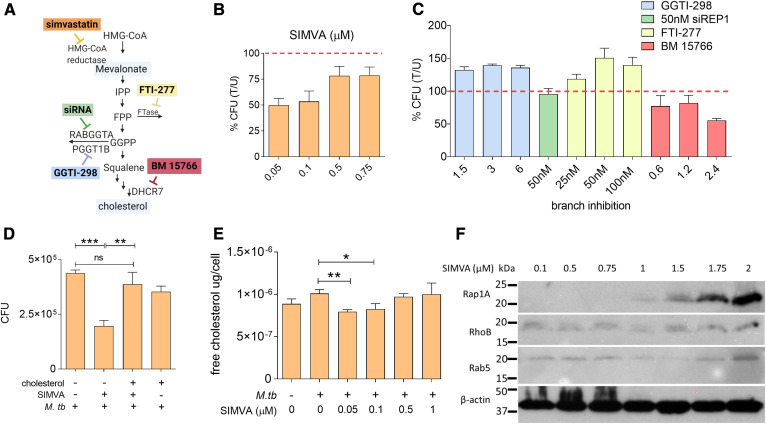
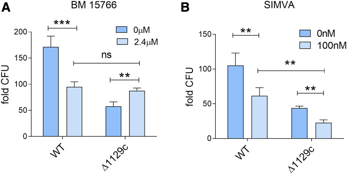
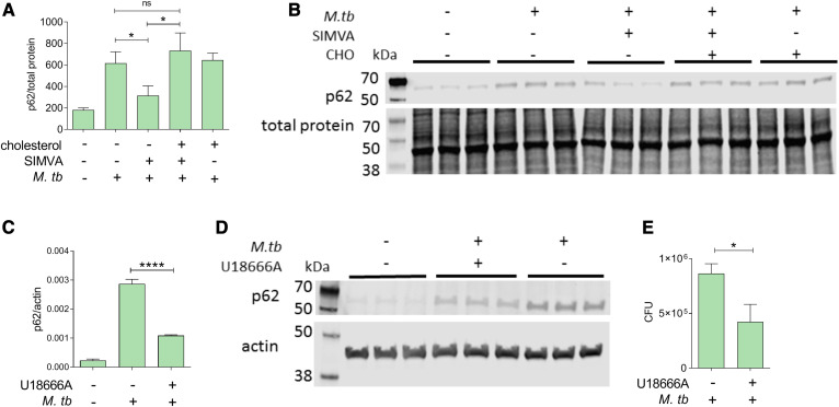
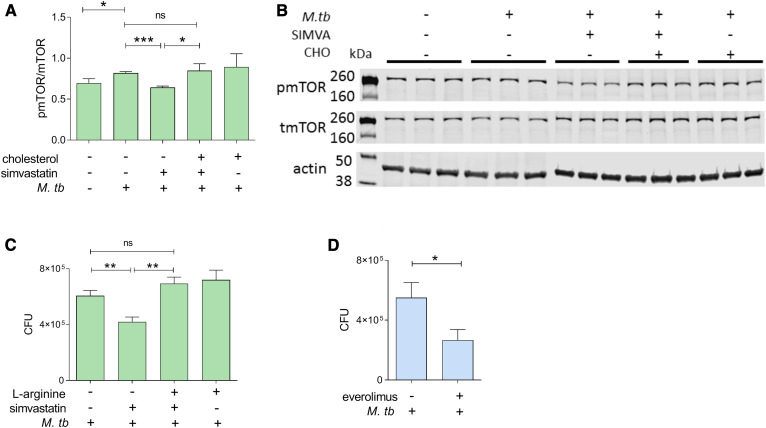
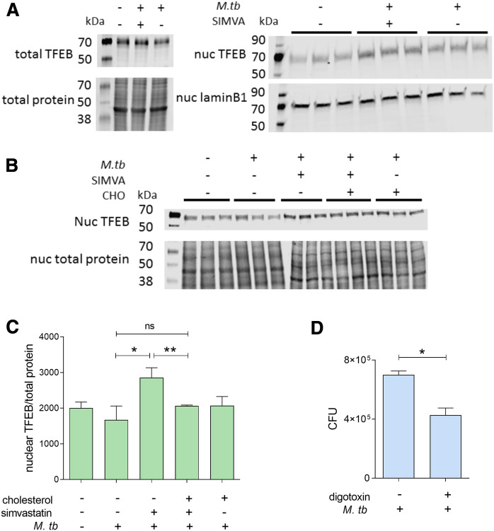
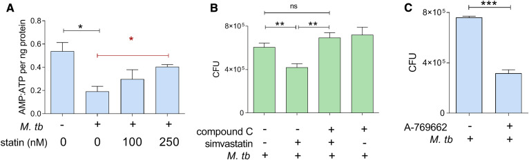
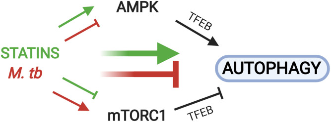
The rise of drug-resistant tuberculosis poses a major risk to public health. Statins, which inhibit both cholesterol biosynthesis and protein prenylation branches of the mevalonate pathway, increase anti-tubercular antibiotic efficacy in animal models. However, the underlying molecular mechanisms are unknown. In this study, we used an in vitro macrophage infection model to investigate simvastatin's anti-tubercular activity by systematically inhibiting each branch of the mevalonate pathway and evaluating the effects of the branch-specific inhibitors on mycobacterial growth. The anti-tubercular activity of simvastatin used at clinically relevant doses specifically targeted the cholesterol biosynthetic branch rather than the prenylation branches of the mevalonate pathway. Using Western blot analysis and AMP/ATP measurements, we found that simvastatin treatment blocked activation of mechanistic target of rapamycin complex 1 (mTORC1), activated AMP-activated protein kinase (AMPK) through increased intracellular AMP:ATP ratios, and favored nuclear translocation of transcription factor EB (TFEB). These mechanisms all induce autophagy, which is anti-mycobacterial. The biological effects of simvastatin on the AMPK-mTORC1-TFEB-autophagy axis were reversed by adding exogenous cholesterol to the cells. Our data demonstrate that the anti-tubercular activity of simvastatin requires inhibiting cholesterol biosynthesis, reveal novel links between cholesterol homeostasis, the AMPK-mTORC1-TFEB axis, and Mycobacterium tuberculosis infection control, and uncover new anti-tubercular therapy targets.

Keywords: Mycobacterium tuberculosis; adenosine 5′-monophosphate-activated protein kinase-mechanistic target of rapamycin complex 1-transcription factor EB axis; immunology; lipids; macrophages/monocytes; mechanistic target of rapamycin complex 1 regulation; statins.


Conflict of interest statement

Conflict of interest—The authors declare that they have no conflicts of interest with the contents of this article.

Figures

Fig. 1.
Fig. 1.
Simvastatin reduces the intracellular burden of M. tuberculosis by inhibiting cellular cholesterol biosynthesis. A: Diagram of the mevalonate pathway. IPP, isopentenyl pyrophosphate; FPP, farnesyl pyrophosphate; FTase, farnesyltransferase; GGPP, geranylgeranyl pyrophosphate; RABGGTA, Rab geranylgeranyltransferase subunit alpha; PGGT1B, protein geranylgeranyltransferase type I subunit beta; DHCR7, 7-dehydrocholesterol reductase. The colored boxes represent pharmacological inhibitors of specific enzymes in the pathway (simvastatin, FTI-277, GGTI-298, BM 15766), as indicated. siRNA targeting REP-1 (a chaperone protein that presents Rab proteins for prenylation by GGTase-II) was used because no pharmacological inhibitor of GGTI type 2 was commercially available. B, C: THP1 cells were infected with M. tuberculosis H37Rv, followed by treatment with multiple doses of simvastatin (B) or branch inhibitors of the mevalonate pathway (C). CFUs were enumerated at 6 days post treatment. Data are presented as percent CFU changes relative to solvent control. U, solvent-treated; T, treated. The red dashed line represents M. tuberculosis CFU levels in the absence of any pharmacological treatment. D: Analysis of cellular cholesterol levels in infected THP1 cells treated with solvent and increasing doses of simvastatin. (E) Intracellular growth of M. tuberculosis in THP1 cells treated for 6 days with 100 nM simvastatin in the absence and presence of water-soluble cholesterol (1.25 μg/ml). Significance was tested by Student’s t-test: *P < 0.05; **P < 0.01 and ***P < 0.001. F: Western blot analysis of sentinel protein targets of the mevalonate pathway of infected THP1 cells treated with simvastatin (SIMVA; 0.1–2 μM) for 6 days. β-Actin was used as loading control. It is noted that, in particular, the antibody against Rap1A is directed against the unprenylated Rap1A form, and therefore, the signal increases with the simvastatin dose.
Fig. 2.
Fig. 2.
Reduction of M. tuberculosis growth by simvastatin is independent of mycobacterial utilization of cholesterol as a carbon source. A, B: THP1 cells were infected with WT or deletion mutant (ΔRv1129c) M. tuberculosis strains for 6 days and treated with either BM 15766 (inhibitor of 7-dehydrocholesterol reductase) (2.4 μM) (A) or simvastatin (100 nM) (B). At day 6 posttreatment, cells were lysed, plated on Middlebrook 7H10 agar plates, and incubated for 3 weeks at 37°C for bacterial CFU enumeration. Data represent fold change in CFU at 6 days relative to 4 h postinfection. Significance was tested by Student’s t-test: **P < 0.01 and ***P < 0.001.
Fig. 3.
Fig. 3.
Reduction of M. tuberculosis growth by simvastatin is associated with cholesterol-dependent induction of autophagy. A, C: Immunoblot analysis of the abundance of p62/SQSTM1 and β-actin or total protein in whole-cell lysates obtained from THP1 cells infected with M. tuberculosis for 6 days and treated with DMSO as a solvent control or 100 nM simvastatin in the absence or presence of water-soluble cholesterol (1.25 μg/ml) (A) and U186666A (inhibitor of cholesterol transport and synthesis) (1.25 μM) (C). Protein quantification and normalization relative to total protein or β-actin per lane was performed using LI-COR Image Studio software. B, D: Representative immunoblots of p62/SQSTM1 in whole-cell lysates of THP1 cells treated with 100 nM simvastatin in the presence of soluble cholesterol (1.25μg/ml) (B) and U186666A (1.25 μM) (D). When total protein was used as loading control (panel B), only a portion of the membrane probed for total protein is shown. E: Effect of U18666A on intracellular growth of M. tuberculosis in THP1 cells. Significance was tested by Student’s t-test: *P < 0.05 and ****P < 0.0001; ns, not significant.
Fig. 4.
Fig. 4.
Reduction of M. tuberculosis growth by simvastatin is associated with cholesterol-dependent reduction of mTORC1 activation. A: Immunoblot analysis of the phosphorylated/total mTORC1 ratio normalized to β-actin in whole-cell lysates of uninfected and infected THP1 cells for 6 days treated with DMSO as a solvent control or 100 nM simvastatin in the absence and presence of soluble cholesterol (1.25 μg/ml). Protein quantification and normalization relative to β-actin per lane was performed using LI-COR Image Studio software. B: A representative image of immunoblots as shown in A. C: Intracellular growth of M. tuberculosis in THP1 cells infected for 6 days and treated with 100 nM simvastatin in the absence or presence of l-arginine (0.78 mM). D: Effect of the mTORC1 inhibitor everolimus on intracellular growth of M. tuberculosis in THP1 cells. Significance was tested by Student’s t-test: *P < 0.05, **P < 0.01 and ***P < 0.001; ns, not significant.
Fig. 5.
Fig. 5.
Reduced M. tuberculosis burden by simvastatin is associated with cholesterol-dependent nuclear translocation of TFEB. A: Representative immunoblots of TFEB in whole-cell lysates (left panel) and nuclear extracts (right panel) of THP1 cells uninfected or infected with M. tuberculosis for 6 days treated with DMSO as a solvent control or 100 nM simvastatin. Protein quantification and normalization relative to total protein or lamin B1, per lane, was performed using LI-COR Image Studio software. B: Immunoblot analysis of TFEB abundance (relative to total protein) in nuclear lysates of infected THP1 cells treated with 100 nM simvastatin in the absence or presence of soluble cholesterol (1.25 μg/ml). When total protein was used as loading control (panels A and B), only a portion of the membrane probed for total protein is shown. C: Effect of 7.5 nM Digoxin (TFEB activator) on the intracellular growth of M. tuberculosis in THP1 cells. Significance was tested by Student’s t-test: *P < 0.05; **P < 0.01; ns, not significant.
Fig. 6.
Fig. 6.
Reduction of M. tuberculosis growth by simvastatin is associated with regulation of the AMPK pathway. A: AMP:ATP ratio in THP1 cells treated with 100 and 250 nM simvastatin, which are both anti-tubercular (supplemental Fig. S3). statin, simvastatin. B: Intracellular growth of M. tuberculosis in THP1 cells infected for 6 days and treated with DMSO as a solvent control or 100 nM simvastatin in the absence or presence of compound C (AMPK inhibitor) (110 nM). C: Effect of 25 μM A-769662 (AMPK activator) on the intracellular growth of M. tuberculosis in THP1 cells. Significance was tested by Student’s t-test (black lines) or by one-way ANOVA test for trend (red line): *P < 0.05; **P < 0.01; ***P < 0.001; ns, not significant.
Fig. 7.
Fig. 7.
Statins and M. tuberculosis infection exhibit opposing effects on AMPK, mTORC1, and autophagy. Reduction of cellular cholesterol content by simvastatin (green arrows) inhibits mTORC1 activation and induces AMPK activation, both of which lead to increased nuclear translocation of TFEB to induce the expression of autophagy-related genes. M. tuberculosis infection (red arrows) induces opposing effects, as it blocks AMPK and induces mTORC1 activation to prevent nuclear translocation of TFEB and block autophagy. For simplicity, the effects of AMPK on mTORC1 activation status and vice versa are not shown.

References

    1. Philips J. A., and Ernst J. D.. 2012. Tuberculosis pathogenesis and immunity. Annu. Rev. Pathol. 7: 353–384. - PubMed
    1. Cadena A. M., Fortune S. M., and Flynn J. L.. 2017. Heterogeneity in tuberculosis. Nat. Rev. Immunol. 17: 691–702. - PMC - PubMed
    1. Karakousis P. C., Yoshimatsu T., Lamichhane G., Woolwine S. C., Nuermberger E. L., Grosset J., and Bishai W. R.. 2004. Dormancy phenotype displayed by extracellular Mycobacterium tuberculosis within artificial granulomas in mice. J. Exp. Med. 200: 647–657. - PMC - PubMed
    1. Lin P. L., Ford C. B., Coleman M. T., Myers A. J., Gawande R., Ioerger T., Sacchettini J., Fortune S. M., and Flynn J. L.. 2014. Sterilization of granulomas is common in active and latent tuberculosis despite within-host variability in bacterial killing. Nat. Med. 20: 75–79. - PMC - PubMed
    1. Gengenbacher M., and Kaufmann S. H. E.. 2012. Mycobacterium tuberculosis: success through dormancy. FEMS Microbiol. Rev. 36: 514–532. - PMC - PubMed

Publication types

MeSH terms

Substances