. 2020 Aug 26;12(558):eaaz5677.
doi: 10.1126/scitranslmed.aaz5677.

A second X chromosome contributes to resilience in a mouse model of Alzheimer's disease

Affiliations

A second X chromosome contributes to resilience in a mouse model of Alzheimer's disease

Emily J Davis et al. Sci Transl Med. .

Abstract

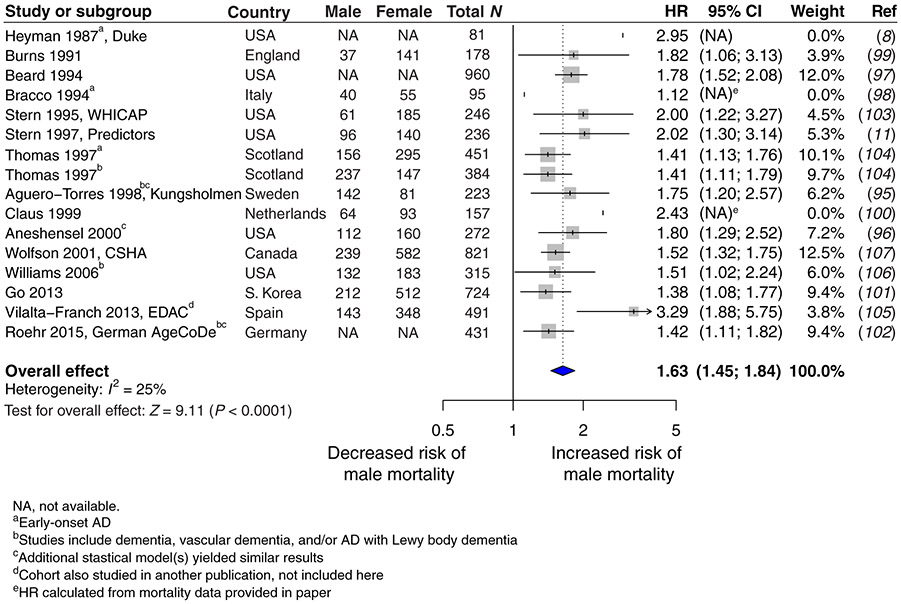
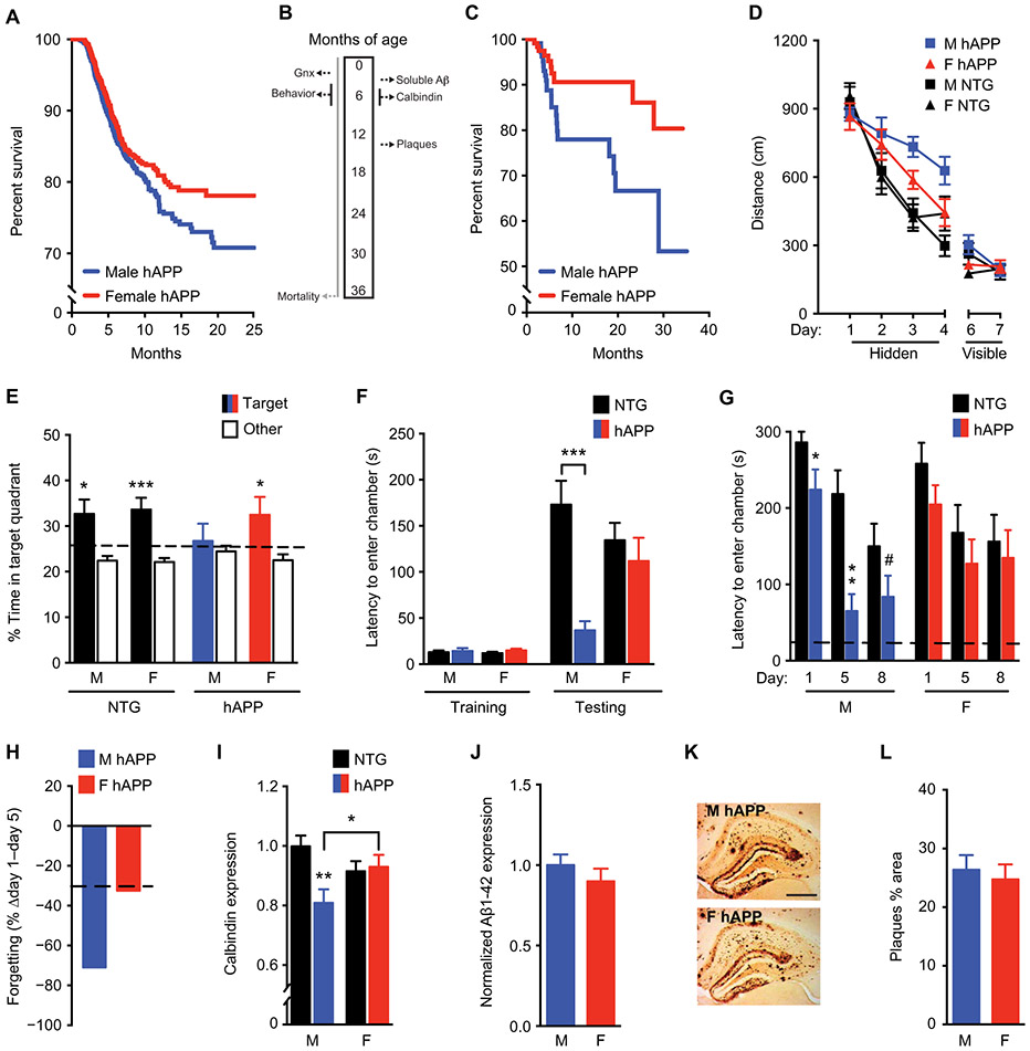
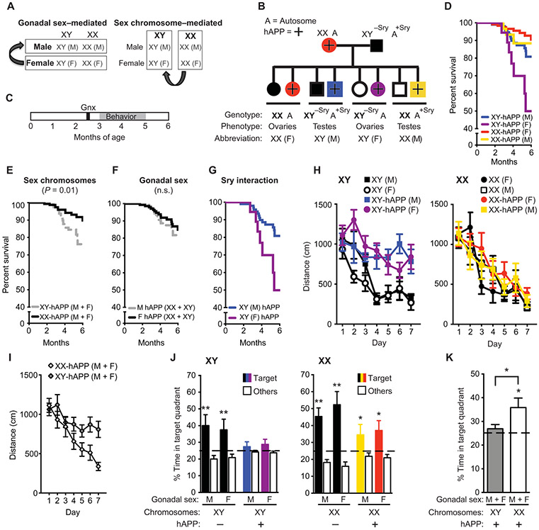
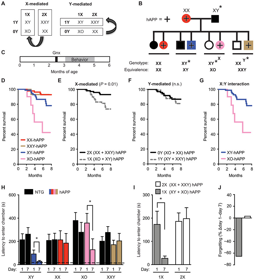
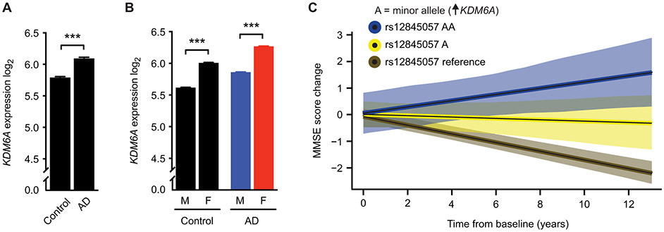
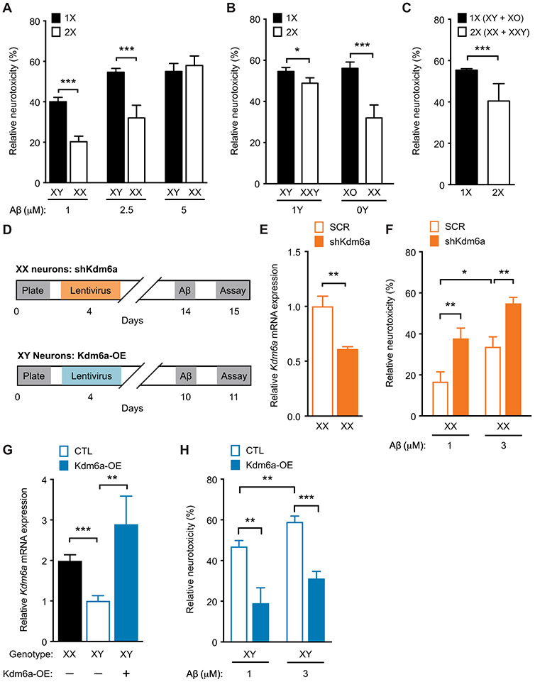
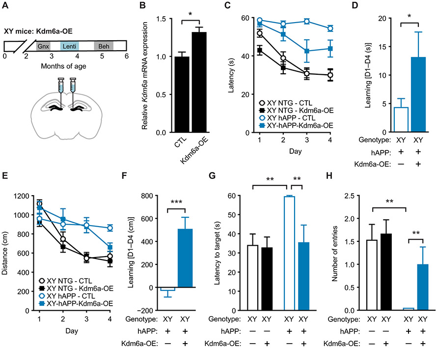
A major sex difference in Alzheimer's disease (AD) is that men with the disease die earlier than do women. In aging and preclinical AD, men also show more cognitive deficits. Here, we show that the X chromosome affects AD-related vulnerability in mice expressing the human amyloid precursor protein (hAPP), a model of AD. XY-hAPP mice genetically modified to develop testicles or ovaries showed worse mortality and deficits than did XX-hAPP mice with either gonad, indicating a sex chromosome effect. To dissect whether the absence of a second X chromosome or the presence of a Y chromosome conferred a disadvantage on male mice, we varied sex chromosome dosage. With or without a Y chromosome, hAPP mice with one X chromosome showed worse mortality and deficits than did those with two X chromosomes. Thus, adding a second X chromosome conferred resilience to XY males and XO females. In addition, the Y chromosome, its sex-determining region Y gene (Sry), or testicular development modified mortality in hAPP mice with one X chromosome such that XY males with testicles survived longer than did XY or XO females with ovaries. Furthermore, a second X chromosome conferred resilience potentially through the candidate gene Kdm6a, which does not undergo X-linked inactivation. In humans, genetic variation in KDM6A was linked to higher brain expression and associated with less cognitive decline in aging and preclinical AD, suggesting its relevance to human brain health. Our study suggests a potential role for sex chromosomes in modulating disease vulnerability related to AD.


Figures

Fig. 1.
Fig. 1.. A meta-analysis of hazard ratios for male and female mortality in AD populations worldwide.
Hazard ratios (HRs) and 95% CIs are shown in a forest plot for studies (8, 11, 95-107) reporting male risk, compared to female risk, for death in longitudinal (and not cross-sectional) analysis of individuals with AD. Overall HR with 95% CI shown in bold indicates increased risk of male mortality (male, HR 1.63, CI 1.45 to 1.84; P < 0.0001). WHICAP, Washington Heights-Inwood Columbia Aging Project; CSHA, Canadian Study of Health and Aging; EDAC, Evolution of Dementia of the Alzheimer-type and Caregiver burden; AgeCoDe, Aging, Cognition, and Dementia in Primary Care Patients.
Fig. 2.
Fig. 2.. Male sex increases mortality, cognitive deficits, and synaptic protein abnormalities in hAPP mice.
(A) Shown are Kaplan-Meier survival curves of male hAPP mice (n = 1572, blue) compared with female hAPP mice (n = 1589, red); all mice had intact gonads (log-rank test, P < 0.001). (B) All mice except those in (A) underwent gonadectomy (Gnx) at about 2.5 months of age; this was followed by behavioral testing conducted from 4 to 7 months of age and survival analysis conducted until 3 years of age. (C) Shown are Kaplan-Meier survival curves of male (n = 116) compared to female (n = 123) hAPP mice after gonadectomy (log-rank test, P < 0.05). (D) Shown are spatial learning curves of mice (age 4 to 7 months; n = 10 to 15 per group) tested in the Morris water maze during hidden platform training and when the platform was visible. Data points are daily average of total distance traveled to reach the platform over four trials. Mixed-model ANOVA for hidden training: female hAPP versus male hAPP mice, P < 0.05. (E) A probe trial was conducted after hidden platform learning and removal of the escape platform. Percentage of time mice spent in the target quadrant of the maze, indicating memory for platform location, versus the average time spent in the other three quadrants is shown; *P < 0.05; ***P < 0.001. The dashed line represents chance performance (25%). (F) Shown is passive avoidance, fear memory of mice (age 3 to 3.5 months; n = 7 to 10 per group) reflected by latency to enter the dark chamber during training and testing 1 day after an electric shock to the foot. Two-way ANOVA: hAPP effect, P < 0.01; hAPP by sex interaction, P < 0.05. (G) Forgetting of passive avoidance memory in a separate cohort of mice (age 5 to 6 months; n = 10 to 12 per group), reflected by latency to enter a dark chamber 1, 5, and 8 days after a foot shock, was measured. The dashed line represents latency to enter the dark chamber during training, which did not differ among groups. (H) Percentage loss of fear memory from days 1 to 5 is shown. The dashed line represents the average for nontransgenic (NTG) animals. (I) Shown is quantitation of calbindin immunoreactivity in mouse dentate gyrus (age 5 to 7 months; n = 11 to 14 mice per group). Two-way ANOVA: hAPP effect, P < 0.05; hAPP by sex interaction, P < 0.05. Means are relative to NTG male control mice, arbitrarily defined as 1. (J) Soluble Aβ1-42 amounts in the mouse hippocampus determined by enzyme-linked immunosorbent assay (ELISA) are shown (age 3 months; n = 8 to 11 mice per group). (K) Representative immunostaining of hippocampal Aβ deposits in coronal brain sections from a male (top, M) and female (bottom, F) hAPP mouse (age 14.5 to 15 months). Scale bar, 200 μm; magnification, ×4. (L) Quantitation of percentage area covered by Aβ deposits in hAPP mice (age 14.5 to 15 months; n = 11 per group). Behavioral studies in male and female NTG and hAPP mice were performed across seven independent cohorts including in fig. S4. #P = 0.06; *P < 0.05; **P < 0.01; ***P < 0.001 [Bonferroni-Holm for (F), (G), and (I)]. Data are presented as means ± SEM.
Fig. 3.
Fig. 3.. Sex chromosomes mediate increased male vulnerability to mortality and cognitive impairments in hAPP mice.
(A) Strategy to identify the cause of sexual dimorphism using the FCG mouse model. (B) Diagram of the cross between hAPP and FCG transgenic mice is presented. FCG mice harbor a transposition of the Sry gene from the Y chromosome onto an autosome (A, autosome). Progeny include XX and XY mice, each with either ovarian (F) or testicular (M) development and with or without hAPP expression (hAPP, +). (C) Experimental strategy: All mice underwent gonadectomy at about 2.5 months of age, followed by behavioral testing and survival studies at 3 to 6 months of age. (D to G) In the Kaplan-Meier survival curves, (D) all groups of hAPP mice showed (E) a main effect of sex chromosomes on mortality (XY, HR 2.49, CI 1.21 to 5.14, P < 0.01) and (F) no main effect of gonadal sex on mortality (P = 0.45). (G) An interaction between sex chromosomes and gonadal sex indicated lower mortality in XY (male, M) compared to XY (female, F) mice (XY-M, HR 0.18, CI 0.03 to 0.92, P < 0.05). Analyses were by Cox proportional hazards for all groups: (XY-M: n = 101; XX-F: n = 122; XY-F: n = 18; XX-M: n = 31). (H and I) Spatial learning curves from the eight genotypes of mice tested altogether in the Morris water maze (age 3 to 5 months; n = 5 to 6 per group) show that (H) XY-hAPP mice (M or F) traveled longer distances to find the target platform, enabling escape from the water maze, than did XX-hAPP mice (M or F). This is highlighted in (I), where all XY-hAPP (M + F) mice were compared with all XX-hAPP (M + F) mice. XX or XY mice without hAPP (M or F) learned similarly well. Data points are daily averages of total distance traveled to reach the platform over four trials. Mixed-model ANOVA: XX-hAPP versus XY-hAPP, P < 0.01. (J and K) A probe trial, during which the escape platform in the target quadrant was removed, tested for memory of the platform location in the eight genotypes of mice. Percentage of time spent in the target quadrant, indicating memory of the platform location, versus the average time spent in the other three quadrants showed that (J) XY-hAPP (M or F) mice did not favor the target quadrant, whereas XX-hAPP (M or F) mice did. The greater impairment of learning and memory in XY-hAPP mice is highlighted in (K) where all XY-hAPP (M + F) mice are compared with all XX-hAPP (M + F) mice. The dashed line represents chance performance. These findings were replicated in an independent cohort (fig. S11). *P < 0.05; **P < 0.01 versus chance performance of 25% (one-sample t tests) or as indicated by bracket (t test). Data are presented as means ± SEM. n.s., not significant.
Fig. 4.
Fig. 4.. A second X chromosome confers resilience against AD-related cognitive impairments in XY (male) and XO (female) hAPP mice.
(A) Strategy to identify whether the sex chromosome effect depends on the X or Y chromosome. (B) Diagram of mouse cross used in this experiment. hAPP females (XX, hAPP) were crossed with XY* males that harbored an altered pseudoautosomal region on the Y chromosome, allowing abnormal crossover with the X chromosome during meiosis (33, 34). The cross resulted in offspring of eight genotypes, each of the sex chromosome genotypes, with or without hAPP. The equivalent number of X and Y chromosomes for each genotype is shown. (C) Experimental strategy: All mice underwent gonadectomy at 2.5 months of age followed by behavioral testing and survival studies between 3 and 6 months of age. (D to G) In the Kaplan-Meier survival curves in (D), all hAPP mice show (E) a main effect of X chromosome dose on mortality (2X, HR 0.2, P < 0.01, CI 0.12 to 0.75) and (F) no main effect of a Y chromosome on mortality (P = 0.53). (G) An interaction between X and Y chromosomes showed lower mortality in the presence of Y (or male gonadal type) when X dose = 1 (XY versus XO, HR 0.23, P < 0.01, CI 0.08 to 0.64). Analyses were by Cox proportional hazards for all groups (XY: n = 79, XX: n = 88; XO: n = 10; XXY: n = 15 mice). (H to J) Shown is testing of mice in the passive avoidance task, measured by latency to enter the dark chamber 1 and 7 days after a foot shock (age 3 to 5 months; n = 4 to 16 per group). (H) Abnormal loss of fear memory in hAPP mice of XY and XO genotypes is shown. Two-way repeated measures ANOVA: X dose effect, P < 0.05. The dashed line represents latency to enter the dark chamber during training, which did not differ among the groups. (I) Greater loss of fear memory in hAPP mice with 1X compared to 2X chromosomes is presented. (J) Percent loss of fear memory in hAPP mice with 1X compared to 2X chromosomes is shown. *P < 0.05 as indicated by bracket (Bonferroni-Holm). Data are presented as means ± SEM.
Fig. 5.
Fig. 5.. A second X chromosome elevates Kdm6a expression independent of gonads or the Y chromosome in mice.
(A) Representative fluorescence in situ hybridization images for Kdm6a and Xist (RNA FISH) expression in XX (top) and XY (bottom) primary mouse neuronal nuclei. Kdm6a is shown in red, Xist is shown in green, and 4’,6-diamidino-2-phenylindole (DAPI) nuclear stain is shown in blue. Nascent Kdm6a transcripts appear as red fluorescent puncta at the site of transcription (indicated by white arrows). Xist RNA remains associated with the inactive X chromosome and is detected only in XX cells. Inset numbers indicate the percentage of nuclei with two sites of nascent Kdm6a accumulation in XX cells and one site in XY cells (n = 100 cells). Scale bar, 2 μm. (B) Representative confocal images of Kdm6a staining (left), Kdm6a with DAPI staining (middle), and Kdm6a with Neuronal nuclei (NeuN) staining (right) in the hippocampal dentate gyrus region of a gonadectomized nontransgenic (NTG) female XX mouse (top row) and a gonadectomized NTG male XY mouse (bottom row). Kdm6a is shown in red, DAPI nuclear stain is shown in blue, and NeuN is shown in green. Scale bar, 50 μm; magnification, ×100. (C and D) Western blot representative image (C) and subsequent quantification (D) of Kdm6a protein expression in the hippocampus of gonadectomized NTG XX female and XY male mice. Bands represent individual mouse samples. (C) Representative images show samples bound by the GeneTex antibody, and (D) quantification is given for both GeneTex and Abcam rabbit anti-Kdm6a antibodies; Kdm6a was normalized using glyceraldehyde phosphate dehydrogenase (GAPDH) as a loading control. Means are relative to NTG XY male control mice, arbitrarily defined as 1 (age 3.4 to 3.6 months; n = 3 mice per group). Gonadectomized NTG XX female mice show higher Kdm6a protein expression. Two-tailed t test, *P < 0.05. (E and F) Hippocampal Kdm6a mRNA expression in (E) FCG mice (age 3.5 to 5.5 months; n = 6 to 26 mice per group) and (F) XY* mice (age 5.5 to 7.5 months; n = 4 to 17 mice per group) with and without hAPP, shown relative to XY male mice without hAPP. Two-way ANOVA: sex chromosome effect, ***P < 0.001 and X dose effect, ***P < 0.001. Data are presented as means ± SEM in (D) to (F). *P < 0.05; ***P < 0.001 (Bonferroni-Holm).
Fig. 6.
Fig. 6.. KDM6A genetic variation associates with cognitive resilience in humans.
(A) Shown is human KDM6A RNA expression via RNA sequencing and microarray in the temporal and parahippocampal cortex of individuals without (control, n = 135) and with AD (n = 86) (***P = 3.64 × 10⁻⁴). (B) Shown is human KDM6A RNA expression via RNA sequencing and microarray in individuals identified as male (M) or female (F) without (M, n = 75; F, n = 60; ***P = 9.79 × 10⁻⁴) and with AD (M, n = 37; F, n = 49; ***P = 4.83 × 10⁻⁴). Expression data were analyzed by linear models accounting for effects of postmortem interval and age at death. (C) Shown is cognitive change with 95% CIs in 778 individuals of the ADNI cohort (cognitively normal, 268; MCI, 465; AD, 45), who carried two alleles (AA, blue, n = 8 all female), one allele (A, yellow, n = 78), or no allele (noncarriers, reference, brown, n = 692) for the rs12845057 variant of the KDM6A gene associated with increased KDM6A RNA expression in brain (table S4). Cognition was measured by the MMSE score. Increasing dose of the minor allele was associated with slower rates of cognitive decline over time (β = 0.141, SE 0.035, P = 0.00005). Cognitive data were analyzed by linear models accounting for effects of baseline age, sex, education, and APOEε4 dose. Data are presented as means ± SEM in (A) and (B). ***P < 0.001 (Bonferroni-Holm).
Fig. 7.
Fig. 7.. Kdm6a knockdown in XX mouse neurons worsens, whereas Kdm6a overexpression in XY neurons attenuates Aβ toxicity in vitro.
(A to C) Vulnerability of mouse primary neurons was tested by the MTT assay. For each genotype, cell toxicity was calculated as a percentage of the corresponding vehicle-treated group, 24 hours after treatment with increasing doses of Aβ. (A) Mouse primary cortical XY neurons showed greater vulnerability than did XX neurons after exposure to vehicle or increasing doses of Aβ (n = 8 to 40 wells per experimental group from 8 to 10 pups per genotype, from four independent litters). Two-way ANOVA: sex chromosome effect, P < 0.01; Aβ dose effect, P < 0.001; interaction, P < 0.05. (B) Toxicity of Aβ in neurons of varying X and Y chromosome dosage derived from littermate pups of XY* males crossed with nontransgenic (NTG) females, with genotypes roughly equivalent to XO, XX, XY, and XX, exposed to vehicle or Aβ (2.5 μM) (n = 15 to 45 wells per experimental group from 7 to 10 pups per genotype, from four independent litters). Two-way ANOVA: X effect, P < 0.0001; Y effect, not significant; X by Y interaction, P < 0.05. (C) Main effect of X chromosome dose shows increased Aβ toxicity in neurons with 1X (XO and XY combined) compared to those with 2X chromosomes (XX and XXY combined). (D) Experimental strategy of lentivirus-mediated knockdown of Kdm6a in XX mouse primary cortical neurons (top) and Kdm6a overexpression in XY mouse primary cortical neurons (bottom). (E) Shown is Kdm6a mRNA expression in neurons transfected with lentivirus expressing scrambled (SCR) or short hairpin (sh) Kdm6a for knockdown expressed relative to XX SCR (n = 5 to 6 wells per experimental group from eight XX pups, from two litters). Two-tailed t test, **P < 0.01. (F) Shown is Aβ toxicity in XX neurons treated with SCR or shKdm6a and exposed to vehicle or Aβ (1 and 3 μM); knockdown of Kdm6a worsened Aβ toxicity (n = 24 to 25 wells per experimental group from 14 XX pups, from three independent litters). Two-way ANOVA: Kdm6a effect, P < 0.001; Aβ effect, P < 0.001; Kdm6a by Aβ interaction, P = 0.99. (G) Kdm6a mRNA expression in neurons transfected with lentivirus expressing control (CTL) or overexpressing Kdm6a (Kdm6a-OE), shown relative to control XY neurons (n = 3 to 8 wells per experimental group from 12 XY pups, from two independent litters). One-way ANOVA, P < 0.001. (H) Shown is Aβ toxicity in XY neurons transfected with lentivirus expressing control or overexpressing Kdm6a (Kdm6a OE) and exposed to vehicle or Aβ (1 and 3 μM); overexpression of Kdm6a attenuated Aβ toxicity (n = 12 to 13 wells per experimental group from 26 XY pups, from three independent litters). Two-way ANOVA: Kdm6a effect, P < 0.001; Aβ effect, P = 0.01; Kdm6a by Aβ interaction, P = 0.99. *P < 0.05; **P < 0.01; ***P < 0.001 (Bonferroni-Holm). Data are presented as means ± SEM.
Fig. 8.
Fig. 8.. Kdm6a overexpression in hippocampus attenuates male vulnerability to cognitive impairments in XY-hAPP mice.
(A) Experimental strategy: XY mice were gonadectomized and injected with lentivirus expressing control or overexpressing Kdm6a (Kdm6a OE) into the dentate gyrus of the hippocampus; animals were then tested on behavioral tasks. (B) Shown is Kdm6a mRNA expression measured in dentate gyrus of mice injected with lentivirus expressing control or overexpressing Kdm6a (Kdm6a OE) (n = 3 mice per experimental group), relative to XY control; t test, *P < 0.05. (C to F) Spatial learning task results for the four experimental groups of XY mice tested in the Morris water maze (age 5 to 5.5 months; n = 7 to 15 per group). XY-hAPP-Kdm6a-OE mice exhibited (C) decreased latency to find the target escape platform (mixed-model ANOVA: XY-hAPP-CTL versus XY-hAPP-Kdm6a-OE, P < 0.001) and (D) a better learning index of latency during hidden platform training, measured by the difference in performance of each mouse at day 4 from average group performance on day 1 (D1 to D4). (E) XY-hAPP-Kdm6a-OE mice did not travel a statistically decreased distance to find the target platform but (F) showed better learning in the distance traveled during hidden platform training. (G and H) Probe trial results 24 hours after completion of hidden platform learning, indicating spatial memory of the escape platform location, showed that XY-hAPP-Kdm6a-OE mice had attenuated spatial deficits including decreased (G) latency to target platform and (H) increased number of entries into the target zone, compared to XY-hAPP-CTL mice. *P < 0.05; **P < 0.01; ***P < 0.001 [Bonferroni-Holm for (G) and (H)]. Data are presented as means ± SEM.

Similar articles

Cited by

References

    1. McCarthy MM, Woolley CS, Arnold AP, Incorporating sex as a biological variable in neuroscience: What do we gain? Nat. Rev. Neurosci 18, 707–708 (2017). - PubMed
    1. Prince M, Wimo A, Guerchet M, Ali G-C, Wu Y-T, Prina M, World Alzheimer Report 2015: The Global Impact of Dementia (Alzheimer’s Disease International (ADI), 2015).
    1. Elmaleh DR, Farlow MR, Conti PS, Tompkins RG, Kundakovic L, Tanzi RE, Developing effective Alzheimer's disease therapies: Clinical experience and future directions. J. Alzheimers Dis 71, 715–732 (2019). - PMC - PubMed
    1. Dubal DB, Sex Differences in Alzheimer’s disease: An updated, balanced, and emerging perspective on differing vulnerabilities, in Sex Differences in Neurology and Psychiatry, Lanzberger R, Kranz GS, Savic-Berglund I, Eds., Handbook of Clinical Neurology, Vol. 175 (3rd series) (Elsevier, 2020); 10.1016/B978-0-444-64123-6.00018-7. - DOI - PubMed
    1. Mielke MM, Vemuri P, Rocca WA, Clinical epidemiology of Alzheimer's disease: Assessing sex and gender differences. Clin. Epidemiol 6, 37–48 (2014). - PMC - PubMed

Publication types