. 2020 Aug 26;10(1):14208.
doi: 10.1038/s41598-020-69783-5.

Nanoscale mechanisms in age-related hip-fractures

Affiliations

Nanoscale mechanisms in age-related hip-fractures

Shaocheng Ma et al. Sci Rep. .

Abstract

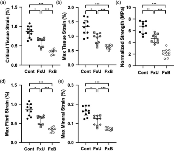
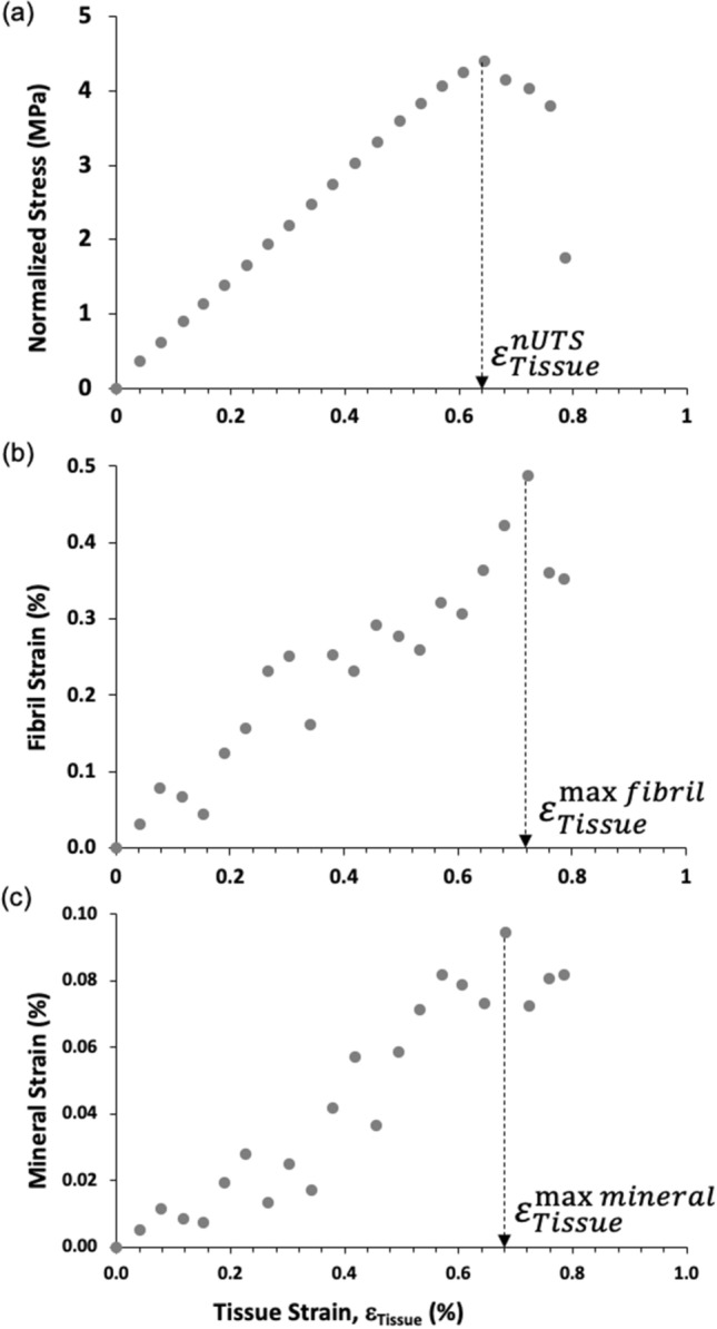
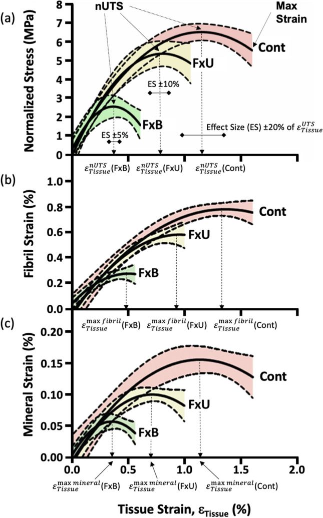
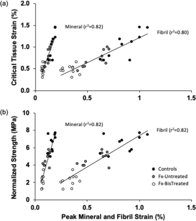
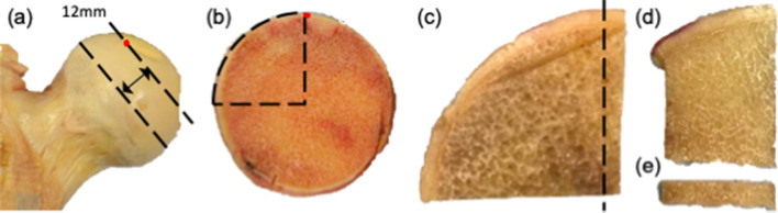
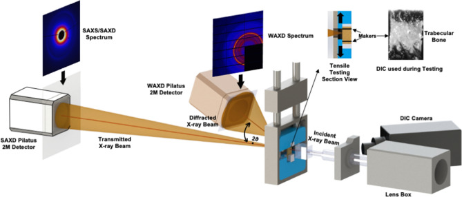
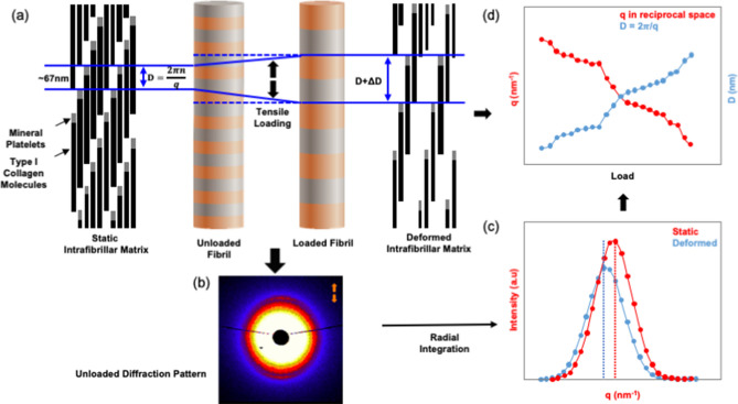
Nanoscale mineralized collagen fibrils may be important determinants of whole-bone mechanical properties and contribute to the risk of age-related fractures. In a cross-sectional study nano- and tissue-level mechanics were compared across trabecular sections from the proximal femora of three groups (n = 10 each): ageing non-fractured donors (Controls); untreated fracture patients (Fx-Untreated); bisphosphonate-treated fracture patients (Fx-BisTreated). Collagen fibril, mineral and tissue mechanics were measured using synchrotron X-Ray diffraction of bone sections under load. Mechanical data were compared across groups, and tissue-level data were regressed against nano. Compared to controls fracture patients exhibited significantly lower critical tissue strain, max strain and normalized strength, with lower peak fibril and mineral strain. Bisphosphonate-treated exhibited the lowest properties. In all three groups, peak mineral strain coincided with maximum tissue strength (i.e. ultimate stress), whilst peak fibril strain occurred afterwards (i.e. higher tissue strain). Tissue strain and strength were positively and strongly correlated with peak fibril and mineral strains. Age-related fractures were associated with lower peak fibril and mineral strain irrespective of treatment. Indicating earlier mineral disengagement and the subsequent onset of fibril sliding is one of the key mechanisms leading to fracture. Treatments for fragility should target collagen-mineral interactions to restore nano-scale strain to that of healthy bone.


Conflict of interest statement

The authors declare no competing interests.

Figures

Figure 1
Figure 1
Comparison of bone tissue and nanomechanics across Control, Fx-Untreated and Fx-BisTreated groups (a) Critical tissue strain (b) Max tissue strain (c) Normalized strength (d) Max fibril strain (e) Max mineral strain. Kruskal Wallis (all p < 0.001) with pairwise Mann–Whitney U tests *p < 0.050, ** < 0.010 and *** < 0.001. Controls (filled black symbols), Fx-Untreated (filled grey symbols), and Fx-BisTreated (open symbols). Females shown as circles and males as squares.
Figure 2
Figure 2
Local displacement curves for a single typical bone sample. (a) Macroscale stress–strain curve indicating the macroscale property of critical tissue strain (εTissuenUTS). (b) Nanoscale fibril-tissue strain curve showing tissue strains to reach the nanoscale property of peak fibril strain (εTissuemaxfibril). (c) Nanoscale mineral-tissue strain curve showing the tissue strains to reach the nanoscale property of peak mineral strain (εTissuemaxmineral). Tissue strain is plotted along the x-axis in all three figures allowing a comparison of the occurrence of key events (the peaks) from the nanoscale to the macroscale.
Figure 3
Figure 3
Overall stress and strain curves for Control, Fx-Untreated and Fx-BisTreated groups. (a) Normalized stress–strain curves indicating the critical (εTissuenUTS) and max tissue strains for each group. Effect sizes (ES) used in the statistical analyses are shown (see Table 2). (b) Fibril-tissue strain curves showing tissue strains to reach peak fibril strain (εTissuemaxfibril). (c) Mineral-tissue strain curves showing the tissue strains to reach peak mineral strain (εTissuemaxmineral). Shaded areas represent the 95% confidence intervals for n = 10 donors per group.
Figure 4
Figure 4
Tissue-level mechanical properties are positively and strongly correlated with peak fibril and mineral strain for both (a) critical tissue strain and (b) normalized strength.
Figure 5
Figure 5
Preparation of bone sections for tensile testing along the axis of the femoral neck (black arrow). (a) Femoral slice cut from the widest point of the femoral head 12 mm thick. (b) Arc sectioned using two more cuts, one along the edge of the fovea capitis (used as a reference point) and one at 90°. (d) Edge piece sectioned from the arc facing fovea and (e) section cut from the end closest to the chiasma along the axis of the femoral neck (12.0 × 2.8 × 1.0 mm).
Figure 6
Figure 6
Schematic diagram of the synchrotron experiment showing both SAXD and WAXD setups to capture X-ray diffraction patterns. A high-speed camera tracked the movement of a tissue sample marked with two black lines for automatically calculating displacement.
Figure 7
Figure 7
Schematic diagram showing fibril deformation and the SAXD method for measuring fibril strain. (a) The 67 nm periodic axial arrangements of type I collagen molecule in the unloaded (i.e. static) and loaded (i.e. deformed) state. D space is the distance between mineral platelets. (b) The corresponding SAXD spectra for the unloaded collagen fibrils as captured by the detector (to the human eye the deformed spectra are imperceptibly different). (c) Corresponding peaks obtained from radial integration of SAXD spectra shows the shift from the static to deformed state. (d) Graph showing how the SAXD diffraction peak shifts after loading and the inverse relationship to the tensile deformation of D-periodic unit in the type I collagen molecule.

References

    1. Rudman K, Aspden R, Meakin J. Compression or tension? The stress distribution in the proximal femur. Biomed. Eng. Online. 2006;5:12. - PMC - PubMed
    1. Yosibash Z, Padan R, Joskowicz L, Milgrom C. A CT-based high-order finite element analysis of the human proximal femur compared to in-vitro experiments. J. Biomech. Eng. 2007;129:297. - PubMed
    1. Bailey AJ, Paul RG, Knott L. Mechanisms of maturation and ageing of collagen. Mech. Ageing Dev. 1998;106:1–56. - PubMed
    1. Burr DB. Bone material properties and mineral matrix contributions to fracture risk or age in women and men. J. Musculoskelet. Neuronal Interact. 2002;2:201–204. - PubMed
    1. Allen MR, Gineyts E, Leeming DJ, Burr DB, Delmas PD. Bisphosphonates alter trabecular bone collagen cross-linking and isomerization in beagle dog vertebra. Osteoporos. Int. 2008;19:329–337. - PubMed

Publication types

Substances