. 2020 Sep;585(7823):129-134.
doi: 10.1038/s41586-020-2646-5. Epub 2020 Aug 26.

Computational design of transmembrane pores

Affiliations

Computational design of transmembrane pores

Chunfu Xu et al. Nature. 2020 Sep.

Abstract

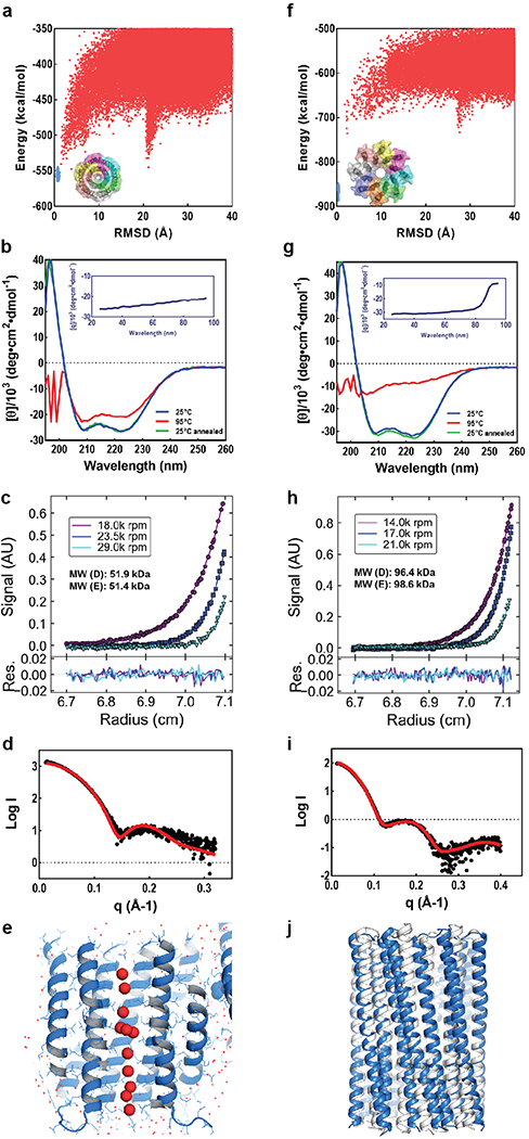
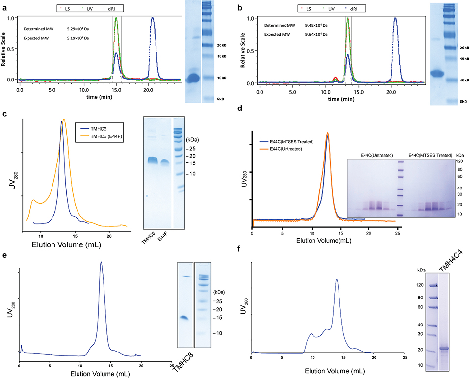
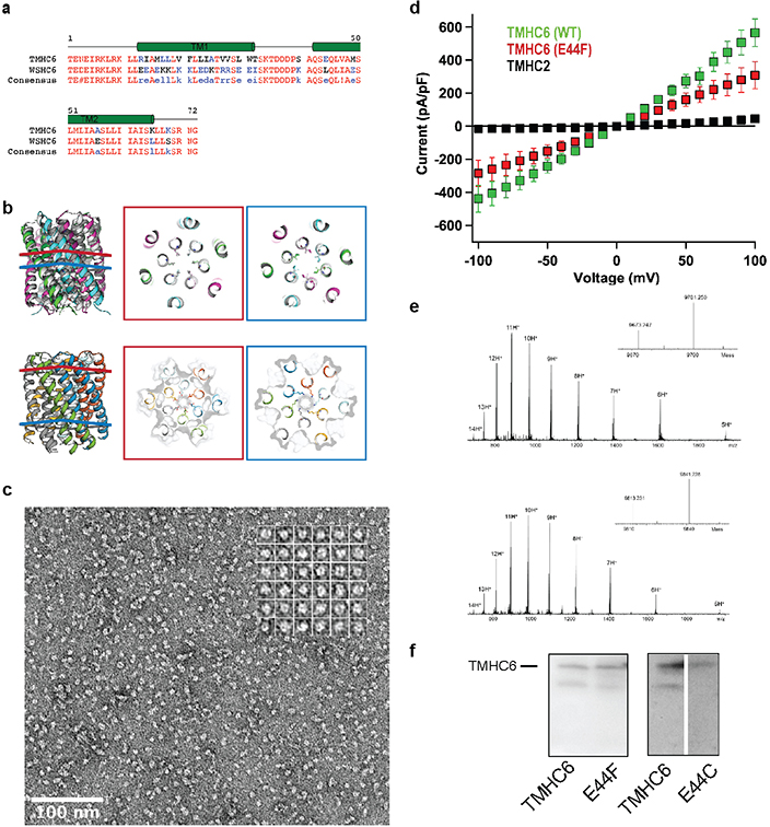
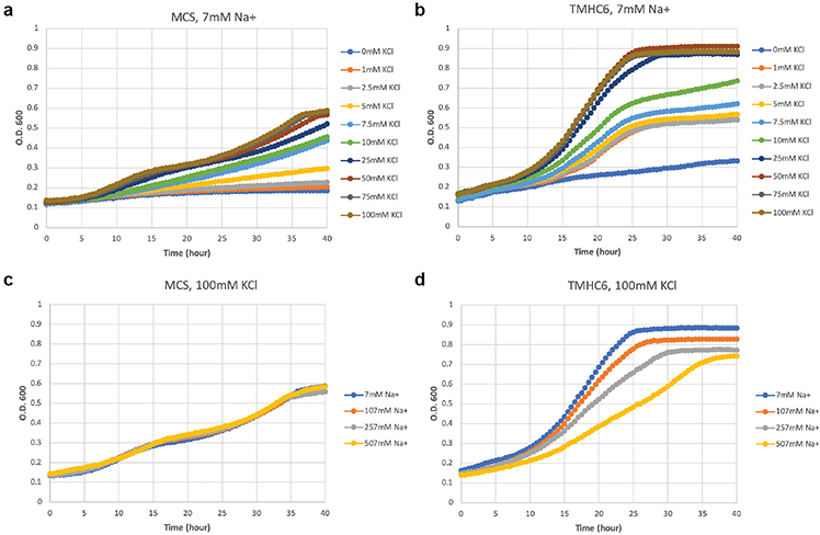
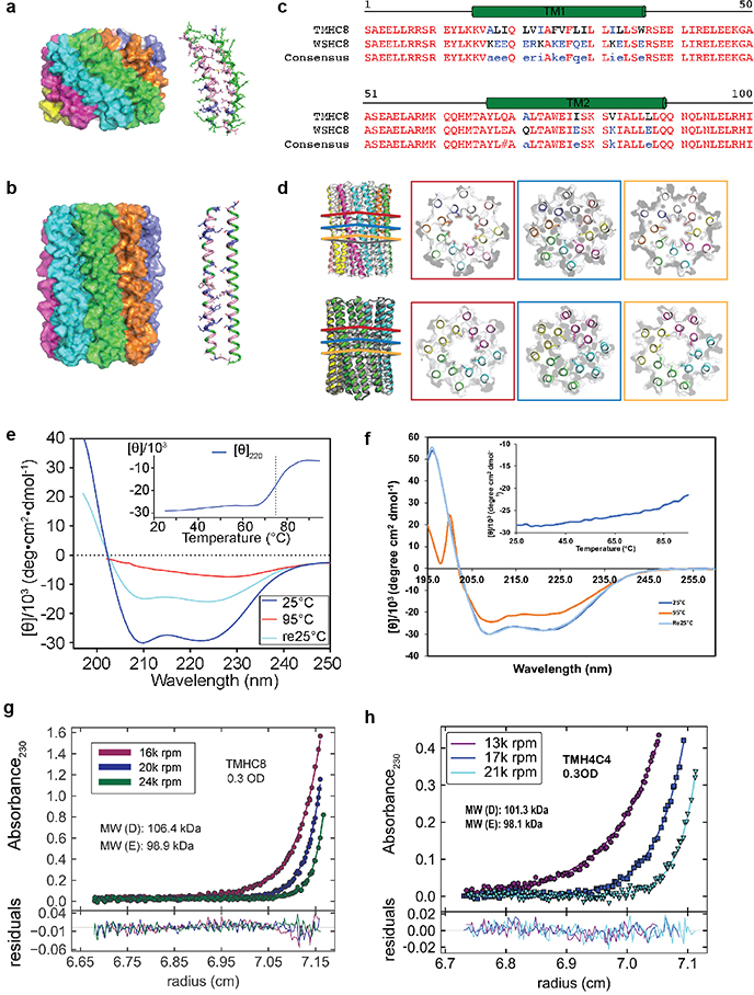
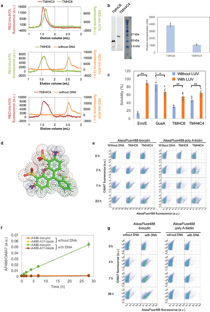
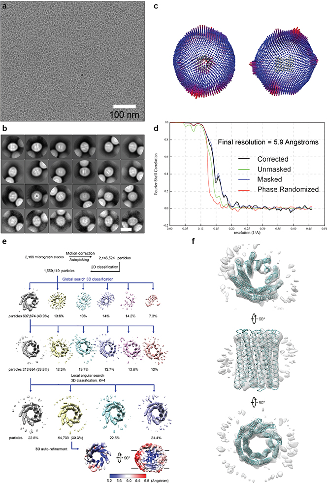
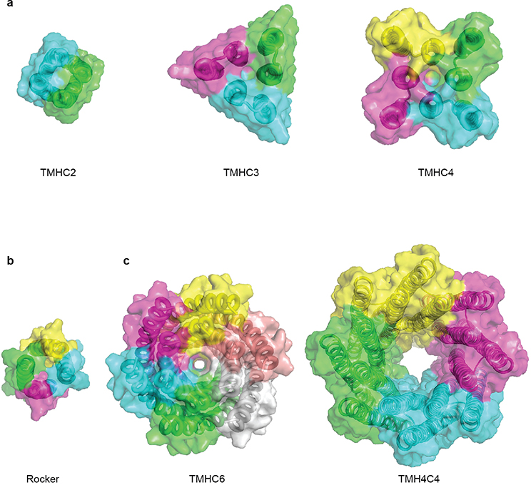
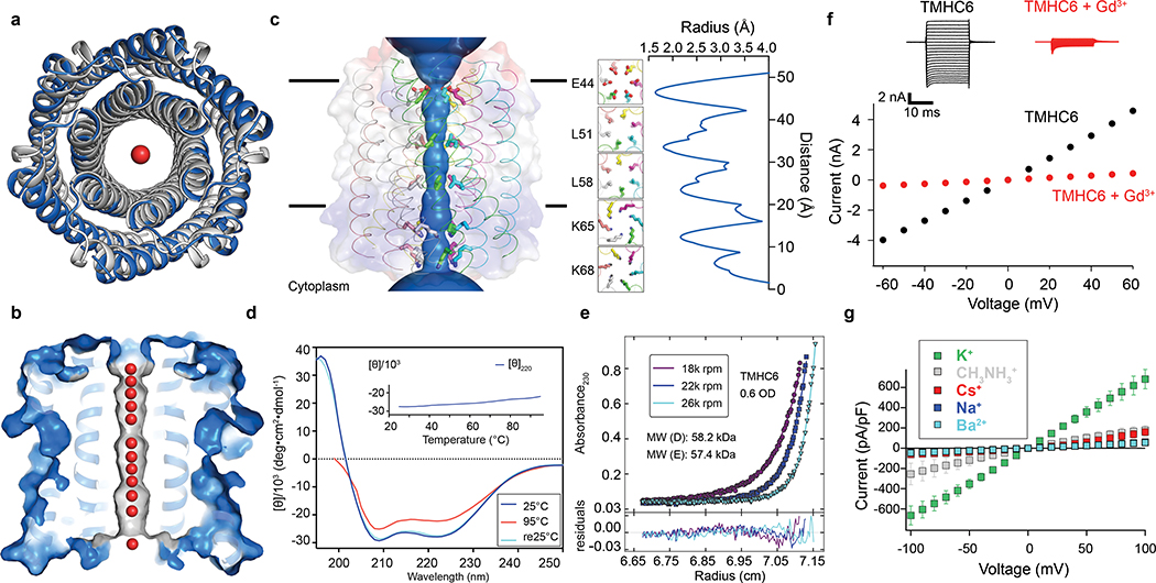
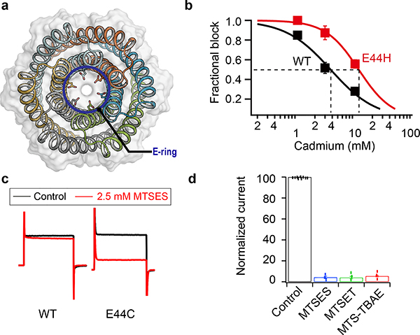
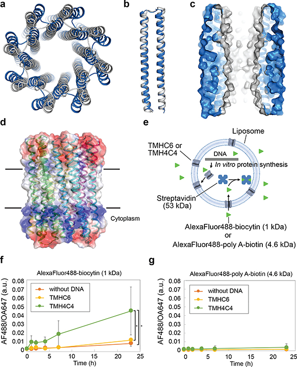
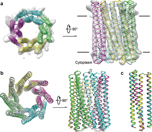
Transmembrane channels and pores have key roles in fundamental biological processes1 and in biotechnological applications such as DNA nanopore sequencing2-4, resulting in considerable interest in the design of pore-containing proteins. Synthetic amphiphilic peptides have been found to form ion channels5,6, and there have been recent advances in de novo membrane protein design7,8 and in redesigning naturally occurring channel-containing proteins9,10. However, the de novo design of stable, well-defined transmembrane protein pores that are capable of conducting ions selectively or are large enough to enable the passage of small-molecule fluorophores remains an outstanding challenge11,12. Here we report the computational design of protein pores formed by two concentric rings of α-helices that are stable and monodisperse in both their water-soluble and their transmembrane forms. Crystal structures of the water-soluble forms of a 12-helical pore and a 16-helical pore closely match the computational design models. Patch-clamp electrophysiology experiments show that, when expressed in insect cells, the transmembrane form of the 12-helix pore enables the passage of ions across the membrane with high selectivity for potassium over sodium; ion passage is blocked by specific chemical modification at the pore entrance. When incorporated into liposomes using in vitro protein synthesis, the transmembrane form of the 16-helix pore-but not the 12-helix pore-enables the passage of biotinylated Alexa Fluor 488. A cryo-electron microscopy structure of the 16-helix transmembrane pore closely matches the design model. The ability to produce structurally and functionally well-defined transmembrane pores opens the door to the creation of designer channels and pores for a wide variety of applications.


Figures

Extended Data Fig. 1 |
Extended Data Fig. 1 |. Design and characterization of water-soluble pores.
(a & f) Design models (insets) and energy versus RMSD plots generated from Rosetta “fold-and-dock” structure prediction calculations. The predicted structures converge on the design models with RMSD values less than 2.0 Å. Structures in the alternative energy minima at large RMSD positions also recapitulate the design models but with chain identities in the RMSD calculations reversed. (b & g) Wavelength-scan and temperature-scan (insets) CD spectra. WSHC6 does not melt up to 95°C, while WSHC8 has a melting temperature of 85°C. The overlap of the pre- and post-annealing CD spectra indicates that the thermal denaturation is reversible. (c & h) Representative analytical ultracentrifugation sedimentation-equilibrium curves at three different rotor speeds for WSHC6 and WSHC8, 0.2 OD230 and 0.3 OD230 in PBS (pH 7.4), respectively. The determined oligomeric states match those of the design models. (d & i) Small-angle X-ray Scattering (SAXS) characterization. The experimental scattering profiles (black) are similar to scattering profiles computed from the design models (red). (e) The chain of water molecules in the pore of WSHC6 crystal structure (red spheres) is verified by processing the data and refining the structure in the P1 space group. (j) Overlay of the crystal structure (blue) and the design model (gray) of WSHC8. Helices are more tilted in the crystal structure than in the design model.
Extended Data Fig. 2 |
Extended Data Fig. 2 |. Representative SEC and SDS-PAGE of the designed proteins.
(a & b) WSHC6 and WSHC8. MWs are determined by coupling SEC with MALS. (c) TMHC6 and E44F mutants. (d) TMHC6 E44C mutant treated or untreated with MTSES reagent. (e) TMHC8. (f) TMH4C4. These experiments were repeated twice with similar results.
Extended Data Fig. 3 |
Extended Data Fig. 3 |. Comparisons between WSHC6 and TMHC6 and additional characterizations of TMHC6 and mutants.
(a) Sequence alignment of TMHC6 with WSHC6. (b) Pore-lining residues in WSHC6 and TMHC6. Top row: overlay of the crystal structure (colors) and the design model (gray) of WSHC6. The pore is lined with alternating leucine (red layer) and isoleucine (blue layer) residues. Bottom row: the TMHC6 pore is lined with E44 ring (red layer) and K65 ring (blue layer) at the extracellular and intracellular sides, respectively. (c) Negative stain EM for TMHC6 in amphipols. Protein particles on the EM grid showed round shape and size consistent with the design model (scale bar at the bottom left, 100 nm). Inset: close-up view of representative particles; each side of the particle frames represent 12.8 nm. (d) Disrupting mutation in the TMHC6 pore entrance reduces the current. The E44F single mutant reduced the K+ current to half of that for TMHC6. TMHC2, a previously designed transmembrane protein without a pore, does not conduct ions across the membrane. 3 cells were measured for each protein and the mean (data points) and standard error of measurement (s.e.m., error bars) are plotted. (e) The covalent modification of TMHC6 E44C mutant by MTSES. Mass-spectrometry results show that there is a 140 Da increase in molecular weight for the mutant after MTSES treatment, in agreement with the predicted value. (f) Expression of TMHC6 and mutants in insect cells for the whole-cell patch-clamp experiments. The same amount of cells were loaded into the gel and the expression levels for two variants were examined by western blot. The E44F mutant had similar, if not higher, expression level as TMHC6. The E44C mutant expressed at a slightly lower level compared to TMHC6. These experiments were repeated three times with similar results.
Extended Data Fig. 4 |
Extended Data Fig. 4 |. The expression of TMHC6 complements a yeast strain defective of K+ uptake.
(a) SGY1528 with an empty vector MCS grew slowly when K+ concentration was lower than 5 mM. Observed growth rate showed dependency on extracellular K+ concentration. (b) SGY1528 supplemented with the TMHC6 gene rescued the yeast growth at lower extracellular K+ concentrations (1 mM - 5 mM) and showed increased growth rates at higher extracellular K+ concentrations (7.5 mM - 100 mM). (c & d) With increasing concentrations of extracellular Na+, TMHC6 yeast showed decreased growth rate (d) in comparison with the insensitive growth rates of MCS yeast (c). These results suggest that TMHC6 conducts K+ and complements the defective K+ uptake in strain SGY1528; this rescuing effect is sensitive to extracellular Na+ concentrations indicating an increased Na+ permeability. A detailed method section is described in Supplementary Materials. The minimal medium and the seeding process are carefully designed to not contain or bring in potassium. The background K+ concentration should be low, which is suggested by the sharp difference between curves for 0 and 1 mM K+ in the TMHC6 case.
Extended Data Fig. 5 |
Extended Data Fig. 5 |. Design and additional characterizations of the designed 16-helix pores.
(a & b) Design models from the first and second rounds of water-soluble designs. The monomers of the first round designs (a, 70 amino acids) are considerably smaller than those of the second round (b, 100 amino acids). (c) Sequence alignment of TMHC8 with WSHC8. (d) Pore-lining residues in WSHC8 and TMH4C4. The crystal and cryoEM structures are in colors. The design models are in gray. Top row: The lumen of WSHC8 pore is freely water-accessible, so the residues inside the pore are all polar. Shown in the figure are three representative layers of the pore-lining residues in the crystal structure, Glu69 ring (red), Lys80 ring (blue), and Glu87 (orange). The missing heavy atoms of these flexible residues are built using Rosetta with backbone constraints. Bottom row: three pore-lining layers in the cryo-EM structure of TMH4C4 corresponding to the three layers in the top row. Glu69 and Glu87 are replaced with glutamine and leucine, respectively. (e & f) CD spectra and thermal stability of 16-helix transmembrane pores. An unfolding transition is observed at ~75°C for TMHC8 (e). TMH4C4 (f) is thermally stable up until 95°C. (g & h) Representative AUC sedimentation-equilibrium curves of 16-helix transmembrane nanopores. By fitting the data globally as a single ideal species in solution, TMHC8 is shown to form complexes with a MW of 98.9 kDa, which is in between the MWs of a heptamer and an octamer. The MW of TMH4C4 is determined to be 98.1 kDa, very close to the MW of a tetramer. ‘MW (D)’ refers to the molecular weight of the oligomer design and ‘MW (E)’ refers to the molecular weight determined in the experiment.
Extended Data Fig. 6 |
Extended Data Fig. 6 |. In vitro protein synthesis and characterizations of TMHC6 and TMH4C4.
(a) SEC analyses of TMHC6 and TMH4C4 purified from E. coli (top) and synthesized in vitro (middle and bottom). (b) The in vitro synthesized products were analyzed by SDS-PAGE and autoradiography51. The means of three independent experiments are shown. The error bars indicate s.e.m.. (c) EmrE, one of the E. coli derived membrane proteins, showed strong interaction with LUV while GusA, soluble enzyme, did not. For TMHC6 and TMH4C4, fraction interacting with LUV was found to be 25.9% and 17.6%, respectively, among synthesized, indicating that the fraction associated with the membrane is similar between them. The mean of four independent experiments are shown. The error bars indicate s.e.m.. Student’s paired t-Test with a two-sided distribution was used to calculate the p values (*p<0.05, **p<0.01; from the left to right p=1.65×10⁻⁶, 0.0357, 0.0040, 0.0024). (d) The narrowest dimension of the head group of Alexa Fluor 488-biocytin is approximately 12 Å. The Van der Waals radius of nitrogen atoms is 1.55 Å. (e) Representative original data for Fig. 3f. Data of approximately 15,000 to 20,000 particles are shown. Similar results were obtained with 7 independent experiments. (f) Flow Cytometry data of the liposomes with pores made of ɑ-hemolysin (AH). Time courses of the median values of the histogram of AF488/OA647 fluorescence, which represents the concentration of the Alexa Fluor 488 inside the liposome are shown. The means of three independent experiments are plotted with the error bars indicating the s.e.m.. (g) Representative original data of (f). Similar results were obtained with 3 independent experiments.
Extended Data Fig. 7 |
Extended Data Fig. 7 |. Cryo-EM resolution estimation and data processing.
(a) Exemplary cryo-EM micrograph of purified TMH4C4 after drift correction and dose-weighting. All the micrographs were with similar results. (b) Class averages after the final round of 2D classification sorted in descending order by the number of particles in each class. The white scale bar in the bottom right panel indicates 10 nm. (c) Angular distribution plot for the final reconstruction from two different views. (d) The gold-standard Fourier shell correlation (FSC) curves for the 3D reconstruction. Deriving map resolution from FSC = 0.143 is indicated. (e) Processing of 2,166 EM micrographs resulted in a total number of 2,146,524 TMH4C4 particles. After a 2D sub-classification, 3D classifications and refinement, a final dataset containing 64,739 particles was used for 3D auto-refinement within Relion 3.0. Local resolution of TMH4C4 was determined within Relion 3.0. Coloured full views (lower lane) from two different orientations illustrate the resolution of different regions in the protein. The low resolution “belt” in the right panel indicates the density for detergents. (f) EM density from 213,654 particles. An EM map (gray) from the second round of 3D classification from 213,654 particles (e) is shown in three perpendicular views. Superposition of the cryo-EM structure of TMH4C4 (cyan) to the map shows a good fit.
Extended Data Fig. 8 |
Extended Data Fig. 8 |. Advances in membrane protein design: from compact helical bundles to transmembrane pores.
(a-b) Surface view of previously reported de novo designed transmembrane proteins,. (c) Surface view of designer transmembrane pores described in this study. Central pores with different sizes are visible.
Figure 1.
Figure 1.. The X-ray crystal structure of the water-soluble hexameric WSHC6 and the ion conductivity of the 12-helix TMHC6 transmembrane channel.
(a) Superposition of backbones of the crystal structure (blue) and the design model (gray) of WSHC6. The C-ɑ RMSD between the crystal structure and the design model is 0.89 Å. (b) The cross section of the WSHC6 channel. A chain of water molecules (red) occupies the central pore (Extended Data Fig. 1e). (c) TMHC6 channel model. The permeation path, calculated by HOLE, is illustrated by the blue surface in the left panel. Constriction sites along the channel are the E-ring (E44), the K-rings (K65, K68), and two intervening L-rings (L51, L58). Right: the radius of the pore along the permeation path. (d) CD spectra and temperature melt (inset) of the TMHC6 channel. No apparent unfolding transition is observed up to 95°C. Re25°C spectrum is taken when the sample cools back to 25°C after the thermal melt scan. (e) AUC sedimentation-equilibrium curves at three different rotor speeds for TMHC6 yield a measured molecular weight of ~58 kDa consistent with the designed hexamer. “MW (D)” and “MW (E)” indicate the molecular weight of the oligomer design and that calculated from the experiment, respectively. (f) Conductivity in whole-cell patch-clamp experiment on insect cells expressing TMHC6. The channel blocker Gd3+ diminished ion conductance to that of untransfected cells. (g) TMHC6 has considerably higher conductance for K+ than Na+, Cs+, CH3NH3+ and Ba2+. 10 cells were measured for each permeant ion species and the mean (data points) and standard error of measurement (s.e.m., error bars) are plotted.
Figure 2.
Figure 2.. Blocking of the ion conductance of TMHC6 with site-directed mutagenesis and chemical biology.
(a) The extracellular ring of six Glu44 residues (E-ring, shown as sticks) is a likely site for cation entry. (b) Cd2+ blocking of the potassium conductance of TMHC6 and the E44H mutant. 3-fold higher Cd2+ concentrations were required to block the E44H mutant compared to the original design, likely because of the reduced electrostatic attraction. 3 cells were measured for each concentration and the mean (data points) and s.e.m. (error bars) are plotted. (c,d) Blocking of the conductance of the TMHC6 E44C mutant using cysteine reactive reagents. In panel (c), y axis shows the current amplitude and x axis indicates the time scale. Negatively charged MTSES, positively charged MTSET, and hydrophobic MTS-TBAE all completely blocked the ion conductance of the E44C mutant within a few minutes under voltage clamp control, while they had no effect on TMHC6 itself in control experiments. 9 cells for the control and 3 cells for each reagent were measured and the bars represent the mean of the measurements.
Figure 3.
Figure 3.. The X-ray crystal structure of water-soluble WSHC8 and the characterization of the 16-helix TMH4C4 transmembrane channel.
(a & b) Superposition of the full octameric assemblies and the monomeric subunits of the crystal structure (blue) and the design model (gray) of WSHC8. The C-ɑ RMSD is 2.51 Å and 0.97 Å, respectively. The larger deviation for the octamer is caused by the slight tilting of the hairpin monomers along the superhelical axis of the complex. (c) The cross section of the WSHC8 channel. (d) TMH4C4 model with 16 transmembrane helices. The electrostatic surface of the neutral transmembrane regions is in gray. (e) Liposome permeability assay. Membrane channels are generated by in vitro translation inside streptavidin containing liposomes, biotin-labeled fluorescent dyes are added to the surrounding buffer, and the amount of dye trapped inside the liposomes is measured by flow cytometry. (f & g) TMH4C4 functions as a size-dependent transmembrane sieve. Incorporation of TMH4C4 into liposomes allowed the accumulation of the 1 kDa but not 4.6 kDa fluorescent dye, while TMHC6 disallowed both. Time courses of the median values of the histogram of Alexa Fluor 488/Ovalbumin conjugated to Alexa Fluor 647 (AF488/OA647) fluorescence (Extended Data Fig. 6e) which represents the concentration of the Alexa Fluor 488 inside the liposome are shown. For (f & g), the error bars indicate s.e.m. (n = 7; the means of the obtained median values are shown). Student’s paired t-Test with a two-sided distribution was used to calculate the p values; *p=0.0128 (right) and 0.0220 (left). In control experiments performed with ɑ-hemolysin (AH) from Staphylococcus aureus, a well-studied channel forming protein with a pore constriction of ~15 Å; only the smaller dye accumulated (Extended Data Fig. 6f–g), suggesting that, as intended, the assay measures solute passage through the transmembrane pores.
Figure 4.
Figure 4.. Cryo-EM of the 16-helix TMH4C4 transmembrane channel.
(a) Cryo-EM density (gray surface) and structure model (colored ribbon) of the 16-helix TMH4C4 protein. EM maps, generated in Chimera, are shown in two perpendicular views. (b) Superposition of the cryo-EM structure (colored) and design model based on the crystal structure of the soluble form (gray) of TMH4C4. (c) Structure alignment of the four protomers in one tetrameric cryo-EM structure of TMH4C4.

References

    1. Gilbert RJC, Bayley H & Anderluh G Membrane pores: from structure and assembly, to medicine and technology. Philos. Trans. R. Soc. Lond. B Biol. Sci 372, (2017). - PMC - PubMed
    1. Eisenstein M An ace in the hole for DNA sequencing. Nature 550, 285–288 (2017). - PubMed
    1. Kasianowicz JJ, Brandin E, Branton D & Deamer DW Characterization of individual polynucleotide molecules using a membrane channel. Proc. Natl. Acad. Sci. U. S. A 93, 13770–13773 (1996). - PMC - PubMed
    1. Clarke J et al. Continuous base identification for single-molecule nanopore DNA sequencing. Nat. Nanotechnol 4, 265–270 (2009). - PubMed
    1. Lear JD, Wasserman ZR & DeGrado WF Synthetic amphiphilic peptide models for protein ion channels. Science 240, 1177–1181 (1988). - PubMed

Publication types

MeSH terms