

Molecular characterization of the 2018 outbreak of lumpy skin disease in cattle in Upper Egypt
- PMID: 32848299
- PMCID: PMC7429391
- DOI: 10.14202/vetworld.2020.1262-1268
Molecular characterization of the 2018 outbreak of lumpy skin disease in cattle in Upper Egypt
Abstract
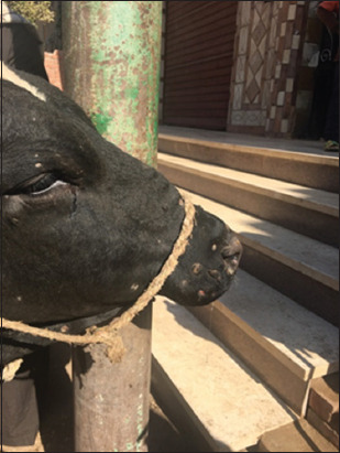
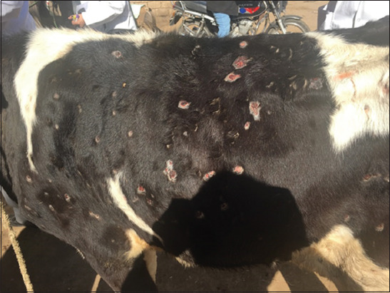
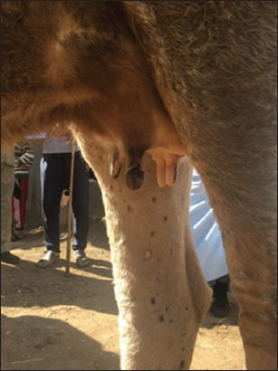
Background and aim: Lumpy skin disease (LSD), an infectious disease of cattle, is characterized by raised nodules on the skin. Although the morbidity rate of LSD is low, it has a considerable fatality rate. Despite the annual mass vaccination of livestock with sheep pox vaccine (Veterinary Serum and Vaccine Research Institute, Egypt) enforced by Egyptian authorities, the LSD virus (LSDV) continues to circulate almost every summer. The present study aimed to discover the cause of cows naturally infected with LSDV circulating in Upper Egypt during the summer of 2018 using polymerase chain reaction (PCR) assay and to analyze their phylogenetics against reference genome sequences.
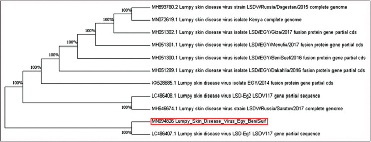
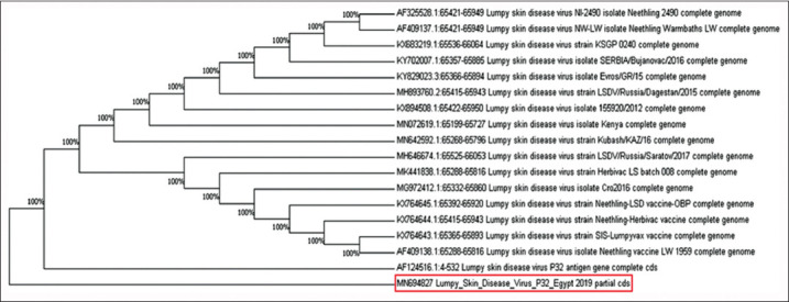
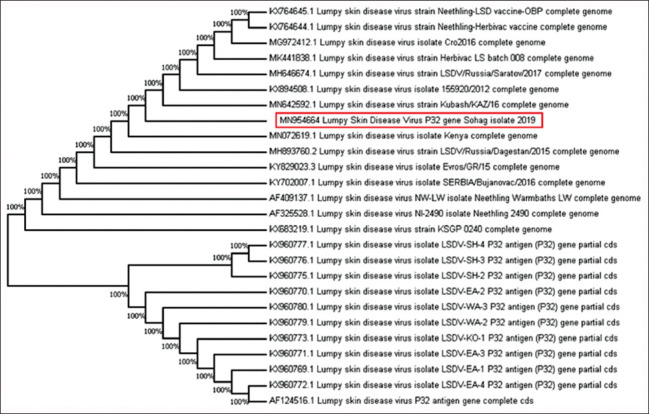
Materials and methods: We cultured LSDV in specific pathogen-free embryonated chicken eggs (SPF-ECE) and used conventional PCR to identify fusion and P32 genes, previously deposited in GenBank (MN694826, MN694827, and MN954664). Sequencing and phylogenetic analyses were performed on these two highly conserved viral genes.
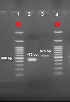
Results: LSDV infection of SPF-ECE resulted in characteristic white pock lesions. PCR products were identified on 1.5% agarose gel after electrophoresis at the expected positions for the fusion and P32 genes at 472 and 587 bp, respectively.
Conclusion: The present study revealed that the two viral genes were identified from the Beni Suef and Sohag Governorates in all clinical cases and confirmed the circulation of LSDV in this outbreak. After sequencing, these genes were identical to those of the LSDV that had been identified and recorded in GenBank for the past 3 years.
Keywords: P32 gene; Upper Egypt; fusion gene; lumpy skin disease; phylogeny.
Copyright: © Allam, et al.
Figures
References
-
- Ahmed A.M, Dessouki A.A. Abattoir-based survey and histopathological findings of lumpy skin disease in cattle at Ismailia abattoir. Int. J. Biosci. Biochem. Bioinform. 2013;3(4):372–375.
-
- Food and Agriculture Organization. Emergence of Lumpy Skin Disease in the Eastern Mediterranean Basin Countries. EMPRES Watch. Vol. 29. Rome: Food and Agriculture Organization; 2013.
-
- Burdin M.L, Prydie J. Lumpy skin disease of cattle in Kenya. Nature. 1959;183(4666):949. - PubMed
-
- Salib F.A, Osman A.H. Incidence of lumpy skin disease among Egyptian cattle in Giza Governorate, Egypt. Vet. (2011) World. 4(4):162–167.
LinkOut - more resources
Full Text Sources
Research Materials
Miscellaneous