

The Role of CTHRC1 in Regulation of Multiple Signaling and Tumor Progression and Metastasis
- PMID: 32848510
- PMCID: PMC7441421
- DOI: 10.1155/2020/9578701
The Role of CTHRC1 in Regulation of Multiple Signaling and Tumor Progression and Metastasis
Abstract
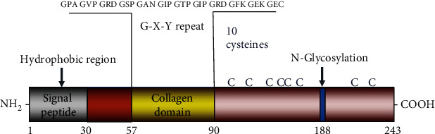
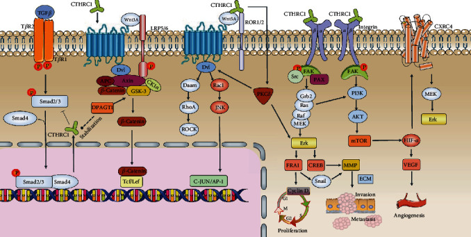
Collagen triple helix repeat containing-1 (CTHRC1) has been identified as cancer-related protein. CTHRC1 expresses mainly in adventitial fibroblasts and neointimal smooth muscle cells of balloon-injured vessels and promotes cell migration and tissue repair in response to injury. CTHRC1 plays a pivotal role in some pathophysiological processes, including increasing bone mass, preventing myelination, and reversing collagen synthesis in many tumor cells. The ascended expression of CTHRC1 is related to tumorigenesis, proliferation, invasion, and metastasis in various human malignancies, including gastric cancer, pancreatic cancer, hepatocellular carcinoma, keloid, breast cancer, colorectal cancer, epithelial ovarian cancer, esophageal squamous cell carcinoma, cervical cancer, non-small-cell lung carcinoma, and melanoma. And molecules that regulate the expression of CTHRC1 include miRNAs, lncRNAs, WAIF1, and DPAGT1. Many reports have pointed that CTHRC1 could exert different effects through several signaling pathways such as TGF-β, Wnt, integrin β/FAK, Src/FAK, MEK/ERK, PI3K/AKT/ERK, HIF-1α, and PKC-δ/ERK signaling pathways. As a participant in tissue remodeling or immune response, CTHRC1 may promote early-stage cancer. Several recent studies have identified CTHRC1 as an effectual prognostic biomarker for predicting tumor recurrence or metastasis. It is worth noting that CTHRC1 has different cellular localization and mechanisms of action in different cells and different microenvironments. In this article, we focus on the advances in the signaling pathways mediated by CTHRC1 in tumors.
Copyright © 2020 Dan Mei et al.
Conflict of interest statement
The authors declare that they have no conflicts of interest.
Figures
Similar articles
-
High expression of Collagen Triple Helix Repeat Containing 1 (CTHRC1) facilitates progression of oesophageal squamous cell carcinoma through MAPK/MEK/ERK/FRA-1 activation.J Exp Clin Cancer Res. 2017 Jun 23;36(1):84. doi: 10.1186/s13046-017-0555-8. J Exp Clin Cancer Res. 2017. PMID: 28645305 Free PMC article.
-
Collagen triple helix repeat containing 1 (CTHRC1) activates Integrin β3/FAK signaling and promotes metastasis in ovarian cancer.J Ovarian Res. 2017 Oct 11;10(1):69. doi: 10.1186/s13048-017-0358-8. J Ovarian Res. 2017. PMID: 29021002 Free PMC article.
-
Collagen triple helix repeat containing-1 negatively regulated by microRNA-30c promotes cell proliferation and metastasis and indicates poor prognosis in breast cancer.J Exp Clin Cancer Res. 2017 Jul 12;36(1):92. doi: 10.1186/s13046-017-0564-7. J Exp Clin Cancer Res. 2017. PMID: 28697793 Free PMC article.
-
Role of collagen triple helix repeat containing-1 in tumor and inflammatory diseases.J Cancer Res Ther. 2017;13(4):621-624. doi: 10.4103/jcrt.JCRT_410_17. J Cancer Res Ther. 2017. PMID: 28901303 Review.
-
The role of collagen triple helix repeat containing 1 (CTHRC1) in cancer development and progression.Expert Opin Ther Targets. 2024 May;28(5):419-435. doi: 10.1080/14728222.2024.2349686. Epub 2024 May 11. Expert Opin Ther Targets. 2024. PMID: 38686865 Free PMC article. Review.
Cited by
-
Collagen Triple Helix Repeat Containing 1 Deficiency Protects Against Airway Remodeling and Inflammation in Asthma Models In Vivo and In Vitro.Inflammation. 2023 Jun;46(3):925-940. doi: 10.1007/s10753-022-01781-3. Epub 2023 Jan 14. Inflammation. 2023. PMID: 36640227
-
Parallel single-cell and bulk transcriptome analyses reveal key features of the gastric tumor microenvironment.Genome Biol. 2022 Dec 22;23(1):265. doi: 10.1186/s13059-022-02828-2. Genome Biol. 2022. PMID: 36550535 Free PMC article.
-
Identification of a tumor microenvironment-related gene signature for predicting prognosis in patients with gastric cancer.Medicine (Baltimore). 2025 Aug 29;104(35):e44032. doi: 10.1097/MD.0000000000044032. Medicine (Baltimore). 2025. PMID: 40898459 Free PMC article.
-
Identification of m6A-associated genes as prognostic and immune-associated biomarkers in Wilms tumor.Discov Oncol. 2023 Nov 8;14(1):201. doi: 10.1007/s12672-023-00817-w. Discov Oncol. 2023. PMID: 37938417 Free PMC article.
-
The whole transcriptome analysis using FFPE and fresh tissue samples identifies the molecular fingerprint of osteosarcoma.Exp Biol Med (Maywood). 2024 Jun 20;249:10161. doi: 10.3389/ebm.2024.10161. eCollection 2024. Exp Biol Med (Maywood). 2024. PMID: 38966281 Free PMC article.
References
-
- Diatchenko L., Lau Y., Campbell A. P., et al. Suppression subtractive hybridization: a method for generating differentially regulated or tissue-specific cDNA probes and libraries. Proceedings of the National Academy of Sciences. 1996;93(12):6025–6030. doi: 10.1073/pnas.93.12.6025. - DOI - PMC - PubMed
Publication types
MeSH terms
Substances
LinkOut - more resources
Full Text Sources
Medical
Research Materials
Miscellaneous
