. 2020 Aug 11:14:780.
doi: 10.3389/fnins.2020.00780. eCollection 2020.

Chromatic Pupillometry Findings in Alzheimer's Disease

Affiliations

Chromatic Pupillometry Findings in Alzheimer's Disease

Martina Romagnoli et al. Front Neurosci. .

Abstract

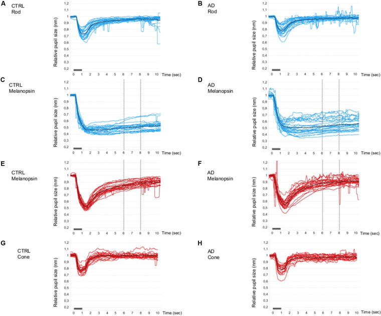
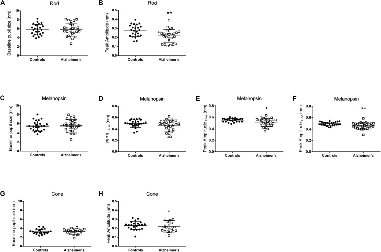
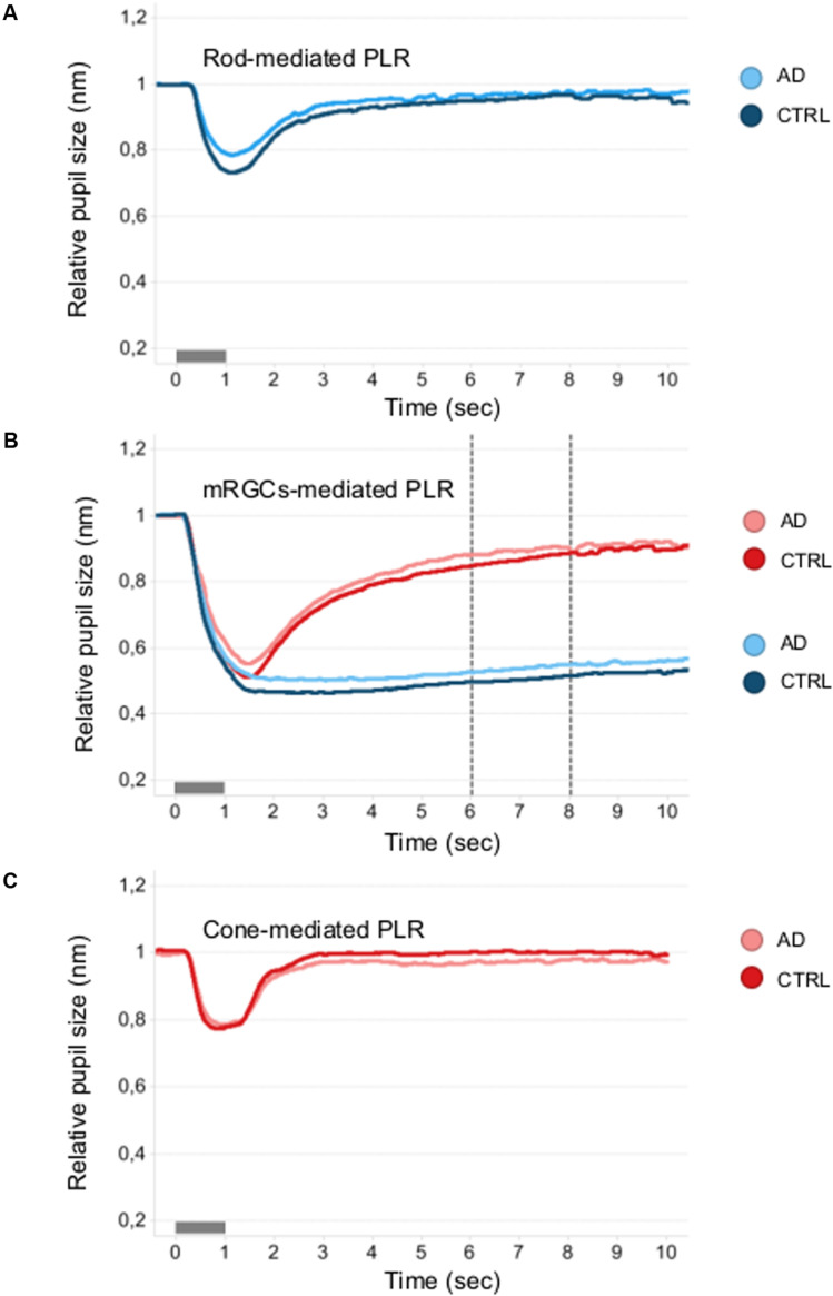
Intrinsically photosensitive melanopsin retinal ganglion cells (mRGCs) are crucial for non-image forming functions of the eye, including the photoentrainment of circadian rhythms and the regulation of the pupillary light reflex (PLR). Chromatic pupillometry, using light stimuli at different wavelengths, makes possible the isolation of the contribution of rods, cones, and mRGCs to the PLR. In particular, post-illumination pupil response (PIPR) is the most reliable pupil metric of mRGC function. We have previously described, in post-mortem investigations of AD retinas, a loss of mRGCs, and in the remaining mRGCs, we demonstrated extensive morphological abnormalities. We noted dendrite varicosities, patchy distribution of melanopsin, and reduced dendrite arborization. In this study, we evaluated, with chromatic pupillometry, the PLR in a cohort of mild-moderate AD patients compared to controls. AD and controls also underwent an extensive ophthalmological evaluation. In our AD cohort, PIPR did not significantly differ from controls, even though we observed a higher variability in the AD group and 5/26 showed PIPR values outside the 2 SD from the control mean values. Moreover, we found a significant difference between AD and controls in terms of rod-mediated transient PLR amplitude. These results suggest that in the early stage of AD there are PLR abnormalities that may reflect a pathology affecting mRGC dendrites before involving the mRGC cell body. Further studies, including AD cases with more severe and longer disease duration, are needed to further explore this hypothesis.

Keywords: Alzheimer’s disease; chromatic pupillometry; melanopsin retinal ganglion cells; post-illumination pupil response; pupil; pupillary light reflex.


Figures

FIGURE 1
FIGURE 1
Single and mean pupillometric waveforms for the rod, melanopsin, and cone conditions in controls and AD. Pupillometric traces obtained under the rod (A,B), melanopsin (C,D, blue and E,F, red), and cone (G,H) conditions of the chromatic pupillometric protocol. Light blue (A–D) and red (E–H) traces represent single individuals, while black traces (A–H) represent the mean waveforms for each group (A,C,E,G for the control group; B,D,F,H for Alzheimer’s group). The vertical dotted lines indicate the time interval (5–7 s from stimulus offset) in which the melanopsin-mediated (sustained) amplitude (PIPR, Post-Illumination Pupil Response, 450 cd/m2) was measured. The light stimulus onset and offset are represented by the gray boxes along the x-axes.
FIGURE 2
FIGURE 2
Pupillometric parameters for controls and AD. Panels show scatterplots with horizontal solid line represents the mean and error bars representing standard deviations for each group. (A,B) Show the results for the Rod-condition; (C–F) Show the results for the Melanopsin-condition [(C–E) 450 cd/m2, 472 nm-blue; (F) 450 cd/m2, 632 nm-red]; (G,H) Show the results for the Cone-condition. (A,C,G) Show normalized pupil size at baseline. (B,E,F,H) Show normalized transient peak amplitude. (D) Show normalized melanopsin-mediated Post-Illumination Pupil Response (PIPRBlue). Peak amplitude (transient peak amplitude) was defined as the difference between the normalized baseline pupil size and the median normalized PLR at the point of maximum pupillary constriction after stimulus onset. PIPR was defined as the difference between the normalized baseline pupil size and the median normalized PLR measured over a 5 to 7 s time interval from stimulus offset. Significant different between controls and AD patients are indicated by an asterix symbol above the groups. *p < 0.05; **p < 0.01.
FIGURE 3
FIGURE 3
Mean pupillometric waveforms obtained under the rod, melanopsin, and cone conditions in controls and AD. (A) Shows PLR measured under the rod-condition with the short- (blue) wavelength flashes (0.001 cd/m2) presented in the dark, for comparison between control (blue mean trace) and AD (azure mean traces) groups. (B) Shows PLR measured under the melanopsin-condition, including the two photopically-matched intense long- (red) and short- (blue) wavelength flashes (450 cd/m2) presented in the dark, for comparison between control (red and blue mean traces) and AD (pink and azure mean traces) groups. The vertical dotted lines indicate the time interval (5–7 s from stimulus offset) over which the melanopsin-mediated (sustained) amplitude (PIPR) was measured. (C) Shows PLR measured with the long- (red) wavelength flashes (10 cd/m2) presented against the rod-suppressing blue adapting field (6 cd/m2) for comparison between control (red mean trace) and AD (pink mean trace) groups.

References

    1. Adhikari P., Pearson C. A., Anderson A. M., Zele A. J., Feigl B. (2015a). Effect of age and refractive error on the melanopsin mediated post-illumination pupil response (PIPR). Sci. Rep. 5:17610. 10.1038/srep17610 - DOI - PMC - PubMed
    1. Adhikari P., Zele A. J., Feigl B. (2015b). The post-Illumination pupil response (PIPR). Invest. Ophthalmol. Vis. Sci. 56 3838–3849. 10.1167/iovs.14-16233 - DOI - PubMed
    1. Baver S. B., Pickard G. E., Sollars P. J., Pickard G. E. (2008). Two types of melanopsin retinal ganglion cell differentially innervate the hypothalamic suprachiasmatic nucleus and the olivary pretectal nucleus. Eur. J. Neurosci. 27 1763–1770. 10.1111/j.1460-9568.2008.06149.x - DOI - PubMed
    1. Beck A. T., Epstein N., Brown G., Steer R. A. (1988). An inventory for measuring clinical anxiety: psychometric properties. J. Consult. Clin. Psychol. 56 893–897. 10.1037//0022-006x.56.6.893 - DOI - PubMed
    1. Beck A. T., Ward C. H., Mendelson M., Mock J., Erbaugh J. (1961). An inventory for measuring depression. Arch. Gen. Psychiatry 4 561–571. 10.1001/archpsyc.1961.01710120031004 - DOI - PubMed