

The Molecular Determinants of Mitochondrial Membrane Contact With ER, Lysosomes and Peroxisomes in Neuronal Physiology and Pathology
- PMID: 32848610
- PMCID: PMC7427582
- DOI: 10.3389/fncel.2020.00194
The Molecular Determinants of Mitochondrial Membrane Contact With ER, Lysosomes and Peroxisomes in Neuronal Physiology and Pathology
Abstract
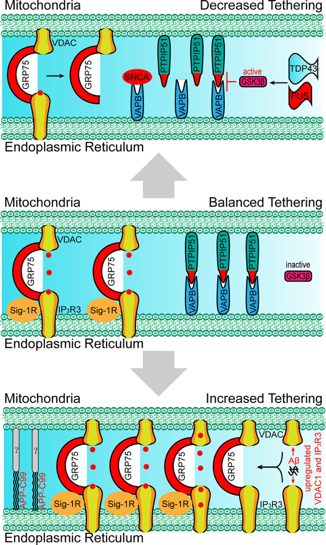
Membrane tethering is an important communication method for membrane-packaged organelles. Mitochondria are organelles with a bilayer membrane, and the membrane contact between mitochondria and other organelles is indispensable for maintaining cellular homeostasis. Increased levels of molecular determinants that mediate the membrane contact between mitochondria and other organelles, and their functions, have been revealed in recent years. In this review article, we aim to summarize the findings on the tethering between mitochondria and other organelles in physiological or pathological conditions, and discuss their roles in cellular homeostasis, neural activity, and neurodegenerative diseases.
Keywords: determinant; membrane contact; mitochondria; neurological diseases; pathological condition; physiological condition.
Copyright © 2020 Liao, Dong and Cheng.
Figures


Similar articles
-
Regulation and Function of Mitochondria-Lysosome Membrane Contact Sites in Cellular Homeostasis.Trends Cell Biol. 2019 Jun;29(6):500-513. doi: 10.1016/j.tcb.2019.02.004. Epub 2019 Mar 18. Trends Cell Biol. 2019. PMID: 30898429 Free PMC article. Review.
-
Peroxisomal Membrane Contact Sites in Mammalian Cells.Front Cell Dev Biol. 2020 Jun 23;8:512. doi: 10.3389/fcell.2020.00512. eCollection 2020. Front Cell Dev Biol. 2020. PMID: 32714927 Free PMC article. Review.
-
Re-evaluation of physical interaction between plant peroxisomes and other organelles using live-cell imaging techniques.J Integr Plant Biol. 2019 Jul;61(7):836-852. doi: 10.1111/jipb.12805. Epub 2019 May 17. J Integr Plant Biol. 2019. PMID: 30916439
-
Unfolded Protein Response-Dependent Communication and Contact among Endoplasmic Reticulum, Mitochondria, and Plasma Membrane.Int J Mol Sci. 2018 Oct 18;19(10):3215. doi: 10.3390/ijms19103215. Int J Mol Sci. 2018. PMID: 30340324 Free PMC article. Review.
-
ER membranes exhibit phase behavior at sites of organelle contact.Proc Natl Acad Sci U S A. 2020 Mar 31;117(13):7225-7235. doi: 10.1073/pnas.1910854117. Epub 2020 Mar 16. Proc Natl Acad Sci U S A. 2020. PMID: 32179693 Free PMC article.
Cited by
-
Cal'MAM'ity at the Endoplasmic Reticulum-Mitochondrial Interface: A Potential Therapeutic Target for Neurodegeneration and Human Immunodeficiency Virus-Associated Neurocognitive Disorders.Front Neurosci. 2021 Oct 21;15:715945. doi: 10.3389/fnins.2021.715945. eCollection 2021. Front Neurosci. 2021. PMID: 34744606 Free PMC article. Review.
-
MERCs. The Novel Assistant to Neurotransmission?Front Neurosci. 2020 Nov 9;14:589319. doi: 10.3389/fnins.2020.589319. eCollection 2020. Front Neurosci. 2020. PMID: 33240039 Free PMC article. Review.
-
The aging of ER-mitochondria communication: A journey from undifferentiated to aged cells.Front Cell Dev Biol. 2022 Aug 19;10:946678. doi: 10.3389/fcell.2022.946678. eCollection 2022. Front Cell Dev Biol. 2022. PMID: 36060801 Free PMC article. Review.
-
Pathological Crosstalk Between Oxidized LDL and ER Stress in Human Diseases: A Comprehensive Review.Front Cell Dev Biol. 2021 May 26;9:674103. doi: 10.3389/fcell.2021.674103. eCollection 2021. Front Cell Dev Biol. 2021. PMID: 34124059 Free PMC article. Review.
References
LinkOut - more resources
Full Text Sources