Review
. 2020 Aug 5:14:208.
doi: 10.3389/fncel.2020.00208. eCollection 2020.

Role of Chondroitin Sulfation Following Spinal Cord Injury

Affiliations
Review

Role of Chondroitin Sulfation Following Spinal Cord Injury

Rowan K Hussein et al. Front Cell Neurosci. .

Abstract

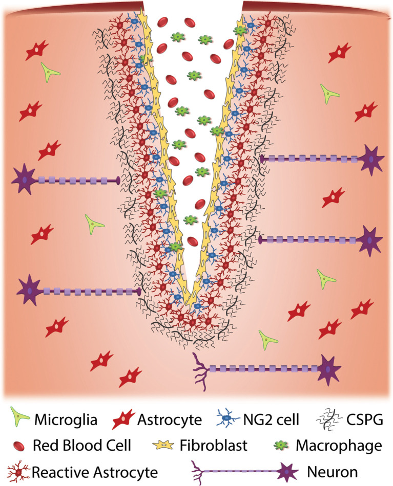
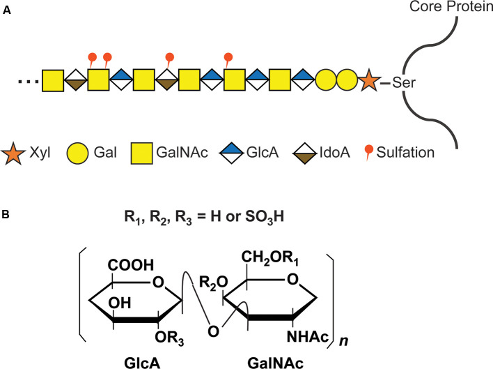
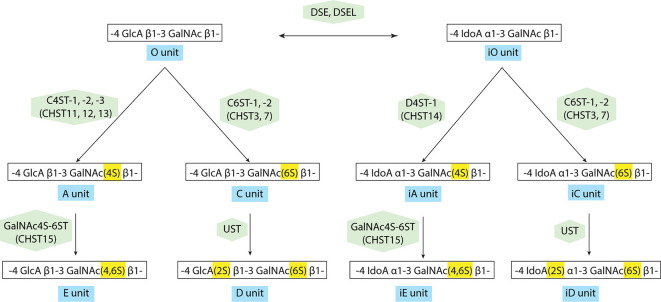
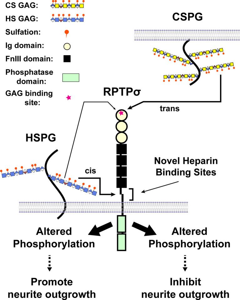
Traumatic spinal cord injury produces long-term neurological damage, and presents a significant public health problem with nearly 18,000 new cases per year in the U.S. The injury results in both acute and chronic changes in the spinal cord, ultimately resulting in the production of a glial scar, consisting of multiple cells including fibroblasts, macrophages, microglia, and reactive astrocytes. Within the scar, there is an accumulation of extracellular matrix (ECM) molecules-primarily tenascins and chondroitin sulfate proteoglycans (CSPGs)-which are considered to be inhibitory to axonal regeneration. In this review article, we discuss the role of CSPGs in the injury response, especially how sulfated glycosaminoglycan (GAG) chains act to inhibit plasticity and regeneration. This includes how sulfation of GAG chains influences their biological activity and interactions with potential receptors. Comprehending the role of CSPGs in the inhibitory properties of the glial scar provides critical knowledge in the much-needed production of new therapies.

Keywords: axon guidance; glial scar; glycosaminoglycan; proteoglycan; receptor tyrosine phosphatase.


Figures

Figure 1
Figure 1
Cellular response to traumatic injury to the central nervous system (CNS). Injury to the CNS results in a series of events that result in the formation of a glial scar around the site of injury. Injury often results in the exposure of nervous system tissue to red blood cells and infiltration from macrophages and fibroblasts. Fibroblasts proliferate at the injury site and eventually form a fibrotic scar. Cellular damage due to injury and exposure to blood initiates an immune response that results in the activation of glial cells such as NG2 cells and astrocytes at the injury site. When activated, these cells produce extracellular matrix (ECM), specifically chondroitin sulfate proteoglycans (CSPGs), at an accelerated rate. The upregulation of CSPGs at the injury site is inhibitory to regenerative axons resulting in a cessation of axonal growth and the formation of retraction bulbs.
Figure 2
Figure 2
CS glycosaminoglycan (GAG) structure and modification by sulfation. (A) Schematic structure of CS GAG chains. CS-GAG chains are attached to the serine residues on the core protein via a tetrasaccharide linkage, followed by the addition of the repetitive disaccharide units modified by different sulfations. (B) Chemical structure of CS disaccharide. CS disaccharides are modified by sulfation at C-4 or C-6 position of GalNAC and/or C-2 position of GlcA.
Figure 3
Figure 3
Modification pathway of CS/dermatan sulfate (DS) GAG chains. After the formation of CS GAG backbone, sulfotransferases transfer sulfate groups from 3′-phosphoadenosine 5′-phosphosulfate (PAPS) to the corresponding positions of GlcA and GalNAc. DS-epimerases convert GlcA into IdoA by epimerizing the C-5 carboxyl group in the chondroitin precursor, resulting in the formation of the dermatan backbone. D4ST1, distinct from C4ST, transfers a sulfate group from PAPS to the C-4 position of the GalNAc residues in dermatan to form the iA-units.
Figure 4
Figure 4
A model illustrating CS and heparan sulfate (HS) binding to RPTPσ. When CS/DS GAG chains are presented by different cells in trans, binding occurs through the first Ig domain far from the transmembrane domain and triggers the changes in phosphorylation (after Katagiri et al., 2018).

Similar articles

Cited by

References

    1. Andrews E. M., Richards R. J., Yin F. Q., Viapiano M. S., Jakeman L. B. (2012). Alterations in chondroitin sulfate proteoglycan expression occur both at and far from the site of spinal contusion injury. Exp. Neurol. 235, 174–187. 10.1016/j.expneurol.2011.09.008 - DOI - PMC - PubMed
    1. Aricescu A. R., McKinnell I. W., Halfter W., Stoker A. W. (2002). Heparan sulfate proteoglycans are ligands for receptor protein tyrosine phosphatase σ. Mol. Cell. Biol. 22, 1881–1892. 10.1128/mcb.22.6.1881-1892.2002 - DOI - PMC - PubMed
    1. Bao X., Nishimura S., Mikami T., Yamada S., Itoh N., Sugahara K. (2004). Chondroitin sulfate/dermatan sulfate hybrid chains from embryonic pig brain, which contain a higher proportion of L-iduronic acid than those from adult pig brain, exhibit neuritogenic and growth factor binding activities. J. Biol. Chem. 279, 9765–9776. 10.1074/jbc.m310877200 - DOI - PubMed
    1. Bellver-Landete V., Bretheau F., Mailhot B., Vallières N., Lessard M., Janelle M. E., et al. . (2019). Microglia are an essential component of the neuroprotective scar that forms after spinal cord injury. Nat. Commun. 10:518. 10.1038/s41467-019-08446-0 - DOI - PMC - PubMed
    1. Bomkamp C., Padmanabhan N., Karimi B., Ge Y., Chao J. T., Loewen C. J. R., et al. . (2019). Mechanisms of PTPσ-mediated presynaptic differentiation. Front. Synaptic Neurosci. 11:17. 10.3389/fnsyn.2019.00017 - DOI - PMC - PubMed

LinkOut - more resources