

Establishment of a Novel Fetal Growth Restriction Model and Development of a Stem-Cell Therapy Using Umbilical Cord-Derived Mesenchymal Stromal Cells
- PMID: 32848614
- PMCID: PMC7401876
- DOI: 10.3389/fncel.2020.00212
Establishment of a Novel Fetal Growth Restriction Model and Development of a Stem-Cell Therapy Using Umbilical Cord-Derived Mesenchymal Stromal Cells
Abstract
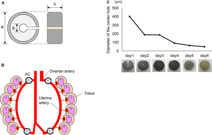
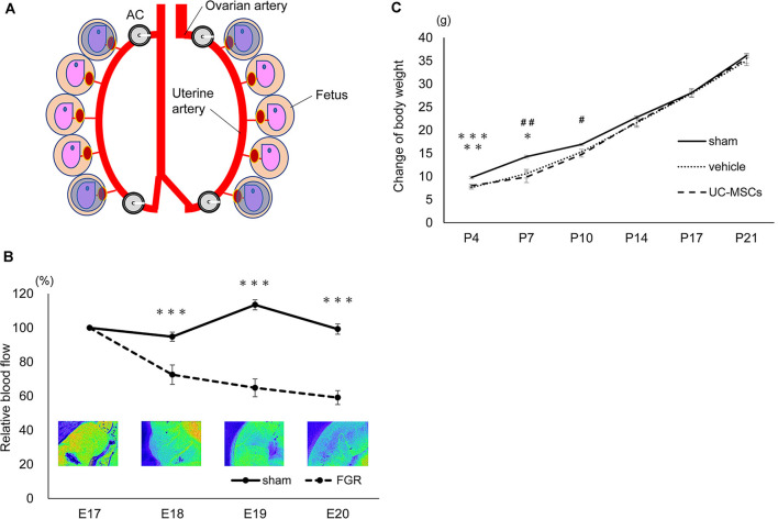
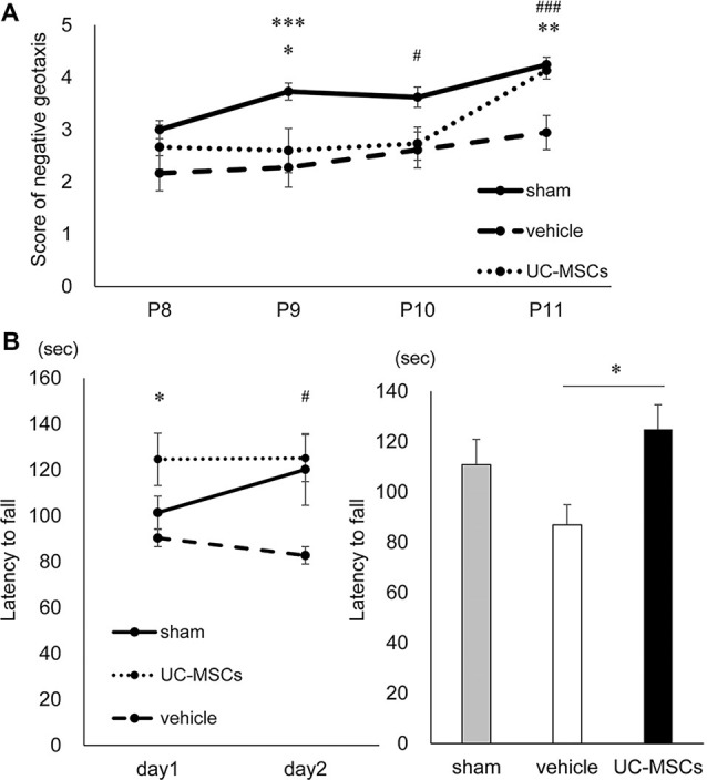
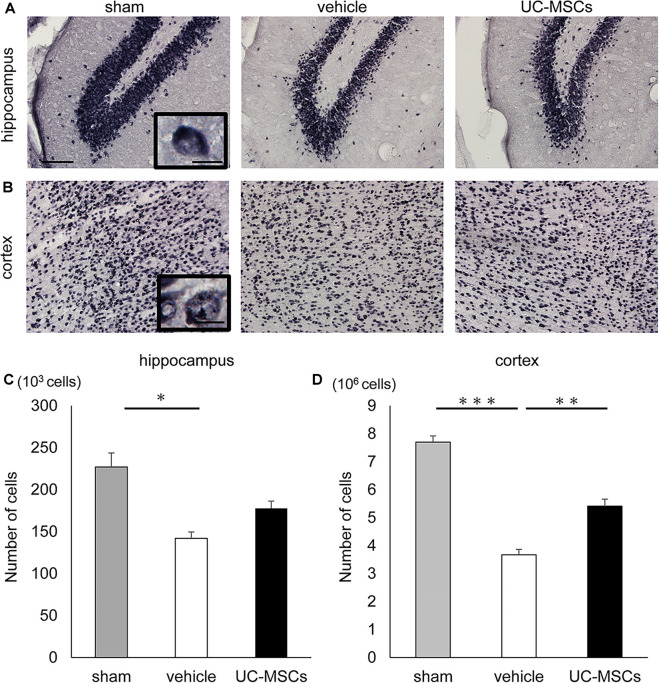
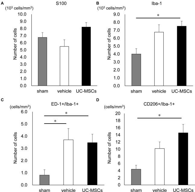
Fetal growth restriction (FGR) is a major complication of prenatal ischemic/hypoxic exposure and affects 5%-10% of pregnancies. It causes various disorders, including neurodevelopmental disabilities due to chronic hypoxia, circulatory failure, and malnutrition via the placenta, and there is no established treatment. Therefore, the development of treatments is an urgent task. We aimed to develop a new FGR rat model with a gradual restrictive load of uterus/placental blood flow and to evaluate the treatment effect of the administration of umbilical cord-derived mesenchymal stromal cells (UC-MSCs). To create the FGR rat model, we used ameroid constrictors that had titanium on the outer wall and were composed of C-shaped casein with a notch and center hole inside that gradually narrowed upon absorbing water. The ameroid constrictors were attached to bilateral ovarian/uterine arteries on the 17th day of pregnancy to induce chronic mild ischemia, which led to FGR with over 20% bodyweight reduction. After the intravenous administration of 1 × 10⁵ UC-MSCs, we confirmed a significant improvement in the UC-MSC group in a negative geotaxis test at 1 week after birth and a rotarod treadmill test at 5 months old. In the immunobiological evaluation, the total number of neurons counted via the stereological counting method was significantly higher in the UC-MSC group than in the vehicle-treated group. These results indicate that the UC-MSCs exerted a treatment effect for neurological impairment in the FGR rats.
Keywords: fetal growth restriction; mesenchymal stem cell; neurodevelopment; stem cells; umbilical cord-derived mesenchymal stromal cells.
Copyright © 2020 Kitase, Sato, Arai, Onoda, Ueda, Go, Mimatsu, Jabary, Suzuki, Ito, Saito, Hirakawa, Mukai, Nagamura-Inoue, Takahashi, Tsuji and Hayakawa.
Figures
Similar articles
-
Human Umbilical Cord-Derived Mesenchymal Stromal Cells Improve Left Ventricular Function, Perfusion, and Remodeling in a Porcine Model of Chronic Myocardial Ischemia.Stem Cells Transl Med. 2016 Aug;5(8):1004-13. doi: 10.5966/sctm.2015-0298. Epub 2016 Jun 22. Stem Cells Transl Med. 2016. PMID: 27334487 Free PMC article.
-
Safety and Efficacy of the Intravenous Infusion of Umbilical Cord Mesenchymal Stem Cells in Patients With Heart Failure: A Phase 1/2 Randomized Controlled Trial (RIMECARD Trial [Randomized Clinical Trial of Intravenous Infusion Umbilical Cord Mesenchymal Stem Cells on Cardiopathy]).Circ Res. 2017 Oct 27;121(10):1192-1204. doi: 10.1161/CIRCRESAHA.117.310712. Epub 2017 Sep 26. Circ Res. 2017. PMID: 28974553 Free PMC article. Clinical Trial.
-
Dose-Dependent Effect of Intravenous Administration of Human Umbilical Cord-Derived Mesenchymal Stem Cells in Neonatal Stroke Mice.Front Neurol. 2018 Mar 8;9:133. doi: 10.3389/fneur.2018.00133. eCollection 2018. Front Neurol. 2018. PMID: 29568282 Free PMC article.
-
Morphometric characteristics of the umbilical cord and vessels in fetal growth restriction and pre-eclampsia.Early Hum Dev. 2016 Jan;92:57-62. doi: 10.1016/j.earlhumdev.2015.11.006. Epub 2015 Dec 8. Early Hum Dev. 2016. PMID: 26678004
-
Neurodevelopment following fetal growth restriction and its relationship with antepartum parameters of placental dysfunction.Ultrasound Obstet Gynecol. 2011 May;37(5):501-14. doi: 10.1002/uog.9008. Ultrasound Obstet Gynecol. 2011. PMID: 21520312 Review.
Cited by
-
Stem Cell Therapy for Neuroprotection in the Growth-Restricted Newborn.Stem Cells Transl Med. 2022 Apr 29;11(4):372-382. doi: 10.1093/stcltm/szac005. Stem Cells Transl Med. 2022. PMID: 35485440 Free PMC article. Review.
-
Combination of human endothelial colony-forming cells and mesenchymal stromal cells exert neuroprotective effects in the growth-restricted newborn.NPJ Regen Med. 2021 Nov 18;6(1):75. doi: 10.1038/s41536-021-00185-5. NPJ Regen Med. 2021. PMID: 34795316 Free PMC article.
-
Altered offspring neurodevelopment in an L-NAME-induced preeclampsia rat model.Front Pediatr. 2023 Jul 13;11:1168173. doi: 10.3389/fped.2023.1168173. eCollection 2023. Front Pediatr. 2023. PMID: 37520045 Free PMC article.
-
Fetal growth restriction followed by early catch-up growth impairs pancreatic islet morphology in male rats.Sci Rep. 2023 Feb 15;13(1):2732. doi: 10.1038/s41598-023-28584-2. Sci Rep. 2023. PMID: 36792668 Free PMC article.
-
Umbilical cord-derived mesenchymal stromal cell therapy to prevent the development of neurodevelopmental disorders related to low birth weight.Sci Rep. 2023 Mar 7;13(1):3841. doi: 10.1038/s41598-023-30817-3. Sci Rep. 2023. PMID: 36882440 Free PMC article.
References
LinkOut - more resources
Full Text Sources
Miscellaneous