. 2020 Jul 30:11:926.
doi: 10.3389/fphys.2020.00926. eCollection 2020.

Chemerin Added to Endothelin-1 Promotes Rat Pulmonary Artery Smooth Muscle Cell Proliferation and Migration

Affiliations

Chemerin Added to Endothelin-1 Promotes Rat Pulmonary Artery Smooth Muscle Cell Proliferation and Migration

Aliénor Hanthazi et al. Front Physiol. .

Abstract

Background: While chemerin has been shown to increase proliferation and migration of systemic vascular smooth muscle cells (SMCs) contributing therefore to the development of hypertension, this remains to be clarified for the pulmonary circulation.

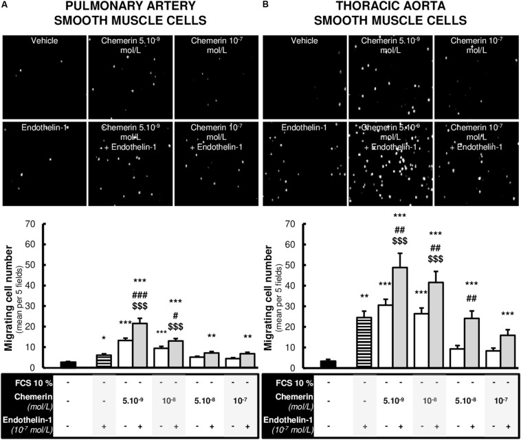
Methods: Expression of chemerin and its three receptors (CMKRL1, CCRL2, GPR1) was examined by immunohistochemistry and RTq-PCR in lungs, pulmonary artery, and thoracic aorta from Wistar rats. Primary cultured rat pulmonary artery and thoracic aorta SMCs treated with recombinant chemerin (tested from 5.10-9 to 10-7 mol/L) were assessed for proliferation and migration (both with 10-7 mol/L endothelin-1), as well as for staurosporine-induced apoptosis.

Results: In pulmonary artery and thoracic aorta, CMKLR1 expression was detected in both endothelial cells and SMCs. In primary cultured pulmonary artery SMCs, chemerin and its three receptors were expressed, and CMKLR1 expression was higher than those of CCRL2 and GPR1. Chemerin added to endothelin-1 increased pulmonary artery SMC proliferation, while chemerin or endothelin-1 alone did not. This effect was less pronounced in thoracic aorta SMCs. Chemerin induced pulmonary artery and thoracic aorta SMC migration, which was exacerbated by endothelin-1 and more pronounced in thoracic aorta SMCs. Chemerin concentration-dependently reduced staurosporine-induced apoptosis in both pulmonary artery and thoracic aorta SMCs. In pulmonary artery SMCs, endothelin-1 treatment increased the expression of CMKLR1, CCRL2, and GPR1, while these expressions were not altered in thoracic aorta SMCs.

Conclusion: Chemerin/CMKRL1 signaling, in conjunction with a key mediator in the pathogenesis of pulmonary hypertensive diseases, endothelin-1, stimulated proliferation and migration, and increased resistance to apoptosis in rat primary cultured pulmonary artery SMCs. Our results suggest that this signaling could play a role in pulmonary artery remodeling observed in pulmonary hypertension.

Keywords: CMKRL1; apoptosis; chemerin; endothelin-1; migration; proliferation; pulmonary artery; smooth muscle cells.


Figures

FIGURE 1
FIGURE 1
Localization of CMKLR1 in rat pulmonary artery and thoracic aorta. Representative images of double immunofluorescent labeling of CMKLR1 (green) with the smooth muscle-specific marker [red, (A)] and with the endothelial-specific marker CD31 [red, (B)] in lungs, as well as in pulmonary artery and thoracic aorta for a total of six fields randomly chosen along each type of tissue from Wistar rats (n = 4) (both positive for elastic fibers observed in green). Negative control images resulted from the overlay of red, green, and blue (DAPI) channels. Labeling in the vascular intimal, medial, and adventitial layers was, respectively, indicated by arrows, #, and $ in both types of vessels. Scale bars = 50 μm for the lung and 25 μm for the pulmonary artery and thoracic aorta sections.
FIGURE 2
FIGURE 2
Expressions of chemerin and its receptors in rat pulmonary artery and thoracic aorta. Relative gene expression of chemerin (A) and its three receptors (B), the chemokine-like receptor 1 (CMKLR1 also called ChemR23; white bars), the C–C chemokine-like receptor 2 (CCRL2; gray bars), and the G protein-coupled receptor 1 (GPR1; black bars) in primary cultured pulmonary artery (seven experiments in total) and thoracic aorta (eight experiments in total) smooth muscle cells isolated from Wistar rats (n = 5). Quantification of relative gene expression was achieved by real-time quantitative polymerase chain reaction (RTq-PCR) using the Pfaffl method with normalization with the housekeeping genes, GAPDH and HPRT1. Results were expressed as relative fold increase over the mean value of relative mRNA expression of chemerin (A) and CMKLR1 (B) in pulmonary artery smooth muscle cells arbitrarily fixed to 1 and presented as mean ± SEM. **0.001 < p < 0.01, ***p < 0.001 compared to the expression of the same gene in pulmonary artery; $0.01 < p < 0.05 compared to the gene expression of CMKLR1 in the same type of vessel.
FIGURE 3
FIGURE 3
Combined chemerin and endothelin-1 treatment increased the proliferation of pulmonary artery smooth muscle cells. Quantification of 5-bromo-2’-deoxyuridine (BrdU) incorporation in primary cultured pulmonary artery [(A), six experiments in total] and thoracic aorta [(B), seven experiments in total] smooth muscle cells isolated from Wistar rats (n = 4), treated or not with endothelin-1 (10–7 mol/L) with increasing concentrations of recombinant mouse chemerin (tested from 5.10–9 to 10–7 mol/L) for 24 h. The proliferative responses were expressed as relative fold increase over the mean value of the proliferative rate of basal condition [corresponding to 0% fetal calf serum (FCS)-treated] in the same type of smooth muscle cells. Results were presented as mean ± SEM. *0.01 < p < 0.05 and ***p < 0.001 compared to corresponding 0% FCS-treated cells; #0.01 < p < 0.05 and ###p < 0.001 compared to corresponding chemerin-treated cells; $$0.001 < p < 0.01 compared to corresponding endothelin-1-treated cells.
FIGURE 4
FIGURE 4
Chemerin decreased the activation of apoptosis in pulmonary artery and thoracic aorta smooth muscle cells. Relative gene expressions of Bax, Bcl2, and pro-apoptotic Bax-to-Bcl2 ratio in primary cultured pulmonary artery [(A,C). six experiments in total] and thoracic aorta [(B,D), seven experiments in total] smooth muscle cells isolated from Wistar rats (n = 5), treated with increasing concentrations of recombinant mouse chemerin (tested from 5.10–9 to 10–7 mol/L). Results were expressed as mean ± SEM. *0.01 < p < 0.05 and **0.001 < p < 0.01 compared to vehicle [0% fetal calf serum (FCS)]-treated smooth muscle cells in the same type of vessels. Representative FACS dot plots and quantification of annexin V/propidium iodide dual labeling in primary cultured pulmonary artery [(E), 14 experiments in total] and thoracic aorta [(F), 13 experiments in total] smooth muscle cells isolated from Wistar rats (n = 8). After 24-h serum starvation, smooth muscle cells were treated with staurosporine (10–6 mol/L) with or without increasing concentrations of recombinant mouse chemerin (tested from 5.10–9 to 10–7 mol/L) for 24 h. Percentages of apoptotic cells corresponded to percentages of cells in early and late apoptosis. Results were presented as mean ± SEM. *0.01 < p < 0.05, **0.001 < p < 0.01 and ***p < 0.001 compared corresponding vehicle (0% FCS)-treated smooth muscle cells; ###p < 0.001 compared to corresponding staurosporine (10–6 mol/L)-treated smooth muscle cells; $p < 0.05 and $$0.001 < p < 0.01 compared to corresponding chemerin (5.10–9 mol/L) and staurosporine (10–6 mol/L)-treated smooth muscle cells.
FIGURE 5
FIGURE 5
Chemerin-induced migration of pulmonary artery and thoracic aorta smooth muscle cells. Representative contrasted images and quantification of in vitro migration of primary cultured pulmonary artery [(A), 12 experiments in total] and thoracic aorta [(B), 9 experiments in total] smooth muscle cells isolated from Wistar rats (n = 4), in the modified Boyden exposed or not to recombinant rat endothelin-1 (10–7 mol/L) with or without increasing concentrations of recombinant mouse chemerin (tested from 5.10–9 to 10–7 mol/L) for 24 h. Migration responses were expressed as mean number of migrating cells evaluated in five different fields randomly chosen per insert (at 10× magnitude) and presented as mean ± SEM. *0.01 < p < 0.05, **0.001 < p < 0.01, ***p < 0.001 compared to basal condition [corresponding to 0% fetal calf serum (FCS)-treated] in the same type of smooth muscle cells; #0.01 < p < 0.05, ##0.001 < p < 0.01 and ###p < 0.001 compared to corresponding chemerin-treated cells; $$$p < 0.001 compared to corresponding endothelin-1-treated cells.
FIGURE 6
FIGURE 6
Altered expression of chemerin receptors induced by endothelin-1 in pulmonary artery and thoracic aorta smooth muscle cells. Relative gene expression of chemerin receptors, the chemokine-like receptor 1 [(A), CMKLR1 also called ChemR23], the C-C chemokine receptor-like 2 [(B), CCRL2], and the G protein-coupled receptor 1 [(C), GPR1] in primary cultured pulmonary artery (five experiments in total) and thoracic aorta (seven experiments in total) smooth muscle cells isolated from Wistar rats (n = 5). After 24-h serum starvation, smooth muscle cells were treated or not with recombinant rat endothelin-1 (10–7 mol/L) for 5 h. Quantification of relative gene expression was achieved by real-time quantitative polymerase chain reaction (RTq-PCR) using the Pfaffl method with normalization with the housekeeping genes, GAPDH and HPRT1. Results were expressed as relative fold increase over the mean value of relative mRNA expression of the vehicle (0% FCS)-treated group arbitrarily fixed to 1 and presented as mean ± SEM. *0.01 < p < 0.05 compared to corresponding vehicle-treated condition. Relative gene expression of endothelin receptors type A [(D), ETA] and B [(E), ETB] in primary cultured pulmonary artery (six experiments in total) and thoracic aorta (six experiments in total) smooth muscle cells isolated from Wistar rats (n = 5). After 24-h serum starvation, smooth muscle cells were treated with recombinant mouse chemerin (5.10–9 and 10–7 mol/L) for 5 h. Quantification of relative gene expression was achieved by real-time quantitative polymerase chain reaction (RTq-PCR) using the Pfaffl method with normalization with the housekeeping genes, GAPDH and HPRT1. Results were expressed as relative fold increase over the mean value of relative mRNA expression of the vehicle (0% FCS)-treated group arbitrarily fixed to 1 and presented as mean ± SEM. *p < 0.05 compared to corresponding vehicle-treated condition.

Similar articles

Cited by

References

    1. Barnea G., Strapps W., Herrada G., Berman Y., Ong J., Kloss B., et al. (2008). The genetic design of signaling cascades to record receptor activation. Proc. Natl. Acad. Sci. U.S.A. 105 64–69. 10.1073/pnas.0710487105 - DOI - PMC - PubMed
    1. Bondue B., Wittamer V., Parmentier M. (2011). Chemerin and its receptors in leukocyte trafficking, inflammation and metabolism. Cytokine Growth. Factor. Rev. 22 331–338. 10.1016/j.cytogfr.2011.11.004 - DOI - PubMed
    1. Bozaoglu K., Bolton K., McMillan J., Zimmet P., Jowett J., Collier G., et al. (2007). Chemerin is a novel adipokine associated with obesity and metabolic syndrome. Endocrinology 148 4687–4694. 10.1210/en.2007-0175 - DOI - PubMed
    1. Carlino C., Trotta E., Stabile H., Morrone S., Bulla R., Soriani A., et al. (2012). Chemerin regulates NK cell accumulation and endothelial cell morphogenesis in the decidua during early pregnancy. J. Clin. Endocrinol. Metab. 97 3603–3612. 10.1210/jc.2012-1102 - DOI - PMC - PubMed
    1. Eddahibi S., Humbert M., Fadel E., Raffestin B., Darmon M., Capron F., et al. (2001). Serotonin transporter overexpression is responsible for pulmonary artery smooth muscle hyperplasia in primary pulmonary hypertension. J. Clin. Invest. 108 1141–1150. 10.1172/jci200112805 - DOI - PMC - PubMed

LinkOut - more resources