Review
. 2020 Aug 7:11:948.
doi: 10.3389/fphys.2020.00948. eCollection 2020.

Structural and Functional Remodeling of the Brain Vasculature Following Stroke

Affiliations
Review

Structural and Functional Remodeling of the Brain Vasculature Following Stroke

Moises Freitas-Andrade et al. Front Physiol. .

Abstract

Maintenance of cerebral blood vessel integrity and regulation of cerebral blood flow ensure proper brain function. The adult human brain represents only a small portion of the body mass, yet about a quarter of the cardiac output is dedicated to energy consumption by brain cells at rest. Due to a low capacity to store energy, brain health is heavily reliant on a steady supply of oxygen and nutrients from the bloodstream, and is thus particularly vulnerable to stroke. Stroke is a leading cause of disability and mortality worldwide. By transiently or permanently limiting tissue perfusion, stroke alters vascular integrity and function, compromising brain homeostasis and leading to widespread consequences from early-onset motor deficits to long-term cognitive decline. While numerous lines of investigation have been undertaken to develop new pharmacological therapies for stroke, only few advances have been made and most clinical trials have failed. Overall, our understanding of the acute and chronic vascular responses to stroke is insufficient, yet a better comprehension of cerebrovascular remodeling following stroke is an essential prerequisite for developing novel therapeutic options. In this review, we present a comprehensive update on post-stroke cerebrovascular remodeling, an important and growing field in neuroscience, by discussing cellular and molecular mechanisms involved, sex differences, limitations of preclinical research design and future directions.

Keywords: angiogenesis; blood–brain barrier; cerebrovascular; neurovascular unit; stroke; vascular remodeling.


Figures

FIGURE 1
FIGURE 1
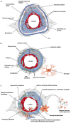
Cellular and acellular constituents of the neurovascular unit (NVU). (A) At the level of penetrating arteries, upstream capillaries, endothelial cells (ECs) are surrounded by vascular smooth muscle cells. At this level, cerebral vessels are still surrounded by the pia. The Virchow–Robin space is located between the pia and the glial limitans formed by the astrocytic endfeet. This perivascular space plays an important role in waste removal and in regulation of the interstitial fluid of the brain. (B) At the level of intracerebral capillaries, the NVU is comprised of ECs, pericytes, astrocytes, microglia, and the basement membrane. Both the ECs and surrounding pericytes are unsheathed by a common basement membrane. Pericyte processes encase most of the endothelial surface. Astrocytic endfeet completely surround the capillary wall. Resting microglial have a ramified morphology and are in constant surveillance around brain microvessels. Gap junction channels enable cytoplasmic continuity between astrocytic endfeet, and also exist between pericytes and ECs at peg-socket structures providing quick communication between these cells. Specialized tight junctions between ECs prevent paracellular leakage into the brain parenchyma. (C) The NVU undergoes dramatic structural changes following stroke, affecting cerebrovascular integrity, neuro-vascular coupling and neuronal survival within the peri-infarct territory. Figure prepared with BioRender.

References

    1. Abbott N. J., Ronnback L., Hansson E. (2006). Astrocyte-endothelial interactions at the blood-brain barrier. Nat. Rev. Neurosci. 7 41–53. 10.1038/nrn1824 - DOI - PubMed
    1. Abdullahi W., Tripathi D., Ronaldson P. T. (2018). Blood-brain barrier dysfunction in ischemic stroke: targeting tight junctions and transporters for vascular protection. Am. J. Physiol. Cell Physiol. 315 C343–C356. - PMC - PubMed
    1. Ahnstedt H., McCullough L. D., Cipolla M. J. (2016). The importance of considering sex differences in translational stroke research. Transl. Stroke Res. 7 261–273. 10.1007/s12975-016-0450-1 - DOI - PMC - PubMed
    1. Al Ahmad A., Taboada C. B., Gassmann M., Ogunshola O. O. (2011). Astrocytes and pericytes differentially modulate blood-brain barrier characteristics during development and hypoxic insult. J. Cereb. Blood Flow Metab. 31 693–705. 10.1038/jcbfm.2010.148 - DOI - PMC - PubMed
    1. Albargothy N. J., Johnston D. A., MacGregor-Sharp M., Weller R. O., Verma A., Hawkes C. A., et al. (2018). Convective influx/glymphatic system: tracers injected into the CSF enter and leave the brain along separate periarterial basement membrane pathways. Acta Neuropathol. 136 139–152. 10.1007/s00401-018-1862-7 - DOI - PMC - PubMed

LinkOut - more resources