. 2020 Aug 4:11:989.
doi: 10.3389/fphys.2020.00989. eCollection 2020.

The Enigma of Endothelium in COVID-19

Affiliations

The Enigma of Endothelium in COVID-19

Savneet Kaur et al. Front Physiol. .

Abstract

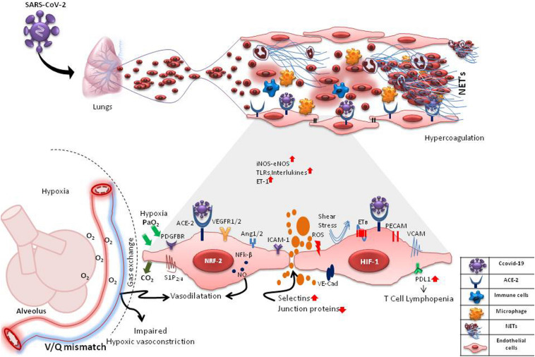
Coronavirus disease 2019 (COVID-19), caused by severe acute respiratory syndrome-related coronavirus-2 (SARS-CoV-2) has affected millions of people globally. Clinically, it presents with mild flu-like symptoms in most cases but can cause respiratory failure in high risk population. With the aim of unearthing newer treatments, scientists all over the globe are striving hard to comprehend the underlying mechanisms of COVID-19. Several studies till date have indicated a dysregulated host immune response as the major cause of COVID-19 induced mortality. In this Perspective, we propose a key role of endothelium, particularly pulmonary endothelium in the pathogenesis of COVID-19. We draw parallels and divergences between COVID-19-induced respiratory distress and bacterial sepsis-induced lung injury and recommend the road ahead with respect to identification of endothelium-based biomarkers and plausible treatments for COVID-19.

Keywords: COVID-19; coagulation; endothelial dysfunction; inflammation; pathogenesis and diagnosis; vascular biology; viral sepsis.


Figures

FIGURE 1
FIGURE 1
Probable Interactions of SARS-CoV-2 with pulmonary microvascular endothelial cells (mECs). SARS-CoV-2 may interact with the mECs directly via ACE2 receptor or indirectly affecting a multitude of endothelial-mediated functions including changes in intercellular permeability, expression of selections, adhesins and VE-cadherins, changes in expression of angiogenic and other functional receptors such as VEGFR1/2, PDGFBR, S1P2/4, PECAMs, ICAMs, VCAM1 causing neutrophilia, formation of neutrophil extracellular traps (NETs), inflammation, hypercoagulation and hypoxia. Hypoxia causes induction of hypoxia-inducible factors (HIF-1) and angiogenesis. ROS from activated neutrophils induces expression of transcription factors, Nrf2 and NFk-B, causing further inflammation. These changes in mECs result in an increase/decrease of vaso-active mediators like nitric oxide (NO), endothelin-1, oxygen sensing machinery, impaired hypoxic vasoconstriction and V/Q mismatch. In inflammatory states, the mECs are also subjected to disturbed blood flow (shear stress). mES dysfunction may also cause an increase in the expression of PD-L1, leading to T cell lymphopenia. These drastic changes in the capillary endothelium disrupts the alveolar-capillary barrier, causing edema and finally respiratory distress.

References

    1. Ackermann M., Verleden S. E., Kuehnel M., Haverich A., Welte T., Laenger F., et al. (2020). Pulmonary vascular endothelialitis, thrombosis, and angiogenesis in Covid-19. N. Engl. J. Med. 383 120–128. 10.1056/nejmoa2015432 - DOI - PMC - PubMed
    1. Aird W. C. (2007). Phenotypic heterogeneity of the endothelium: II. Representative vascular beds. Circul. Res. 100 174–190. 10.1161/01.res.0000255690.03436.ae - DOI - PubMed
    1. Alvarado-Moreno J. A., Majluf-Cruz A. (2020). COVID-19 and dysfunctional endothelium: the mexican scenario. Arch. Med. Res. 10.1016/j.arcmed.2020.05.004 - DOI - PMC - PubMed
    1. Amraei R., Rahimi N. (2020). COVID-19, renin-angiotensin system and endothelial dysfunction. Cells 9:1652. 10.3390/cells9071652 - DOI - PMC - PubMed
    1. Colmenero I., Santonja C., Alonso-Riaño M., Noguera-Morel L., Hernández-Martín A., Andina D., et al. (2020). SARS-CoV-2 endothelial infection causes COVID-19 chilblains: histopathological, immunohistochemical and ultraestructural study of 7 paediatric cases. Br. J. Dermatol.. 10.1111/bjd.19327 - DOI - PMC - PubMed