. 2020 Aug 11:11:993.
doi: 10.3389/fphys.2020.00993. eCollection 2020.

Communication Among Photoreceptors and the Central Clock Affects Sleep Profile

Affiliations

Communication Among Photoreceptors and the Central Clock Affects Sleep Profile

Milena Damulewicz et al. Front Physiol. .

Abstract

Light is one of the most important factors regulating rhythmical behavior of Drosophila melanogaster. It is received by different photoreceptors and entrains the circadian clock, which controls sleep. The retina is known to be essential for light perception, as it is composed of specialized light-sensitive cells which transmit signal to deeper parts of the brain. In this study we examined the role of specific photoreceptor types and peripheral oscillators located in these cells in the regulation of sleep pattern. We showed that sleep is controlled by the visual system in a very complex way. Photoreceptors expressing Rh1, Rh3 are involved in night-time sleep regulation, while cells expressing Rh5 and Rh6 affect sleep both during the day and night. Moreover, Hofbauer-Buchner (HB) eyelets which can directly contact with s-LN v s and l-LN v s play a wake-promoting function during the day. In addition, we showed that L2 interneurons, which receive signal from R1-6, form direct synaptic contacts with l-LN v s, which provides new light input to the clock network.

Keywords: Drosophila; Hofbauer-Buchner eyelets; peripheral clock; photoreceptors; sleep.


Figures

FIGURE 1
FIGURE 1
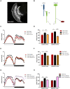
Effects of glass-expressing cells on sleep. (A) Sleep pattern of flies with blocked neurotransmission from photoreceptors (GMR>TeTx) measured as number of sleep bins per hour. (B) Total sleep time of GMR>TeTx flies measured as minutes per 12 h, separately for day and night-time. (C) Sleep pattern of flies with clock disruption in photoreceptors (GMR>Δcyc24). (D) Total sleep time of GMR>Δcyc24 flies. Heterozygous parental strains were used as control. Statistically significant differences marked with asterisk *p ≤ 0.05; **p ≤ 0.01; ****p ≤ 0.0001. Detailed statistics are presented in Supplementary Tables S3, S4.
FIGURE 2
FIGURE 2
Effects of peripheral clocks located in the photoreceptors on the presynaptic protein Bruchpilot (BRP) expression. The immunofluorescence signal intensity was measured in the distal lamina on cryosections of control [Canton S, (A)] and experimental [GMR>Δcyc24, (B)] flies at four time points (ZT1, ZT4, ZT13, and ZT16). Statistically significant differences were marked with letters, where different letters above the bar means confirmed changes between time points. (C) Confocal images of BRP immunostaining in the lamina of GMR>Δcyc24 at different time points.
FIGURE 3
FIGURE 3
Effects of peripheral clock located in the photoreceptors on per expression in the pacemaker. The immunofluorescence signal intensity was measured in s-LNvs and l-LNvs marked with anti-PDF staining for GMR>TeTx (A,B), and GMR>Δcyc24 (C,D) experimental flies. Asterisks (*p ≤ 0.05; **p ≤ 0.01; ****p ≤ 0.0001) show statistically significant differences between experimental flies and controls at specific time point. Detailed statistics are presented in Supplementary Table S5.
FIGURE 4
FIGURE 4
R1-6 photoreceptors control sleep during the night. (A) Rh1-expressing cells are R1-6 photoreceptors, with terminals in the lamina (cryosection of Rh1>GFP brain). (B) Graphical presentation of pathways which are blocked after TeTx expression in Rh1-expressing cells (a – amacrine cells, L1-3 – lamina monopolar cells). (C) Sleep pattern of Rh1>TeTx flies. (D) Total sleep amount during the day and night after blocking of synaptic transmission from R1-6 (Rh1>TeTx). (E) Sleep pattern for Rh1>Δcyc24 flies. (F) Total sleep time of Rh1>Δcyc24 strain. Heterozygous parental strains were used as control. Statistically significant differences marked with asterisks ****p ≤ 0.0001. Detailed statistics are presented in Supplementary Table S3.
FIGURE 5
FIGURE 5
Rh3-expressing cells affect night-time sleep. (A) Rh3 is expressed in R7 and R8 cells, which terminate in the medulla (cryosection of Rh3>GFP brain). (B) Graphical presentation of synaptic connections formed by Rh3-expressing cells which are blocked in Rh3>TeTx flies (Tm – transmedulla neurons). (C) Sleep pattern of Rh3>TeTx flies. (D) Sleep time during the day and night of Rh3>TeTx flies. (E) Sleep pattern of Rh3>Δcyc24. (F) Amount of sleep during the day and night of Rh3>Δcyc24. Heterozygous parental strains were used as control. Statistically significant differences marked with asterisks **p ≤ 0.01; ***p ≤ 0.001; ****p ≤ 0.0001. Detailed statistics are presented in Supplementary Table S3.
FIGURE 6
FIGURE 6
Rh5-expressing cells affect sleep both, during the day and night. (A) Rh5 is expressed in R8 cell which terminates in the medulla (cryosection of Rh5>GFP brain). (B) Graphical representation of pathways blocked in Rh5>TeTx strain. Solid line represents input to R8 cell coming from R7. Dashed line represents blocked output from R8 to downstream cells. (C) Sleep pattern of Rh5>TeTx. (D) Sleep time during the day and night of Rh5>TeTx flies. (E) Sleep pattern of Rh5>Δcyc24. (F) Amount of sleep during the day and night of Rh5>Δcyc24. Heterozygous parental strains were used as control. Statistically significant differences marked with asterisks *p ≤ 0.05; ***p ≤ 0.001; ****p ≤ 0.0001). Detailed statistics are presented in Supplementary Table S3.
FIGURE 7
FIGURE 7
Rh6-expressing cells regulate sleep during the day and night. (A) Rh6-expressing photoreceptors (R8) terminate in the medulla. The cryosection of Rh6>GFP brain does not show Hofbauer-Buchner (HB) eyelets. (B) Graphical presentation of synaptic contacts (dotted line) between Rh6-expressing photoreceptors or HB eyelets and their targets. Solid line represent input to R8 coming from R7 cell. (C) Sleep pattern of Rh6>TeTx flies. (D) Sleep time of Rh6>TeTx flies is increased during the day and night. (E) Sleep pattern of Rh6>Δcyc24. (F) Sleep amount of Rh6>Δcyc24. (G) Sleep pattern of flies with downregulated acetylcholine synthesis in HB eyelets (Rh6>ChAT-RNAi). (H) Sleep amount of Rh6>ChAT-RNAi is increased during the day only. Heterozygous parental strains were used as control, additional control Rh6>Valium10-GFP was used for the last experiment. Statistically significant differences marked with asterisks *p ≤ 0.05; ****p ≤ 0.0001. Detailed statistics are presented in Supplementary Table S3.
FIGURE 8
FIGURE 8
L2 interneurons play important role in the regulation of sleep. (A) Graphical presentation of synaptic contacts formed by L2 (dotted line) (Tm –Transmedulla neurons). (B) L2 terminals are located in close proximity to PDF-immunoreactive LNvs neurons (whole mount immunostaining of L2>GFP flies with anti-GFP and anti-PDF antibodies, blue). (C) Sleep pattern of L2>TeTx flies. (D) Sleep amount presented for flies with tetanus toxin expression in L2 cells. Statistically significant differences marked with asterisks ***p < 0.001; ****p < 0.0001. (E) Fluorescence calcium indicator expressed in L2 cells was measured in the terminals in the medulla. (F) Calcium level in the L2 terminals measured in flies kept in LD12:12 conditions. (G) Calcium level in the L2 terminals of flies in constant darkness (DD). Statistically significant differences were marked with letters, where different letters above the bar means confirmed changes between time points.
FIGURE 9
FIGURE 9
Hofbauer-Buchner eyelets contact with LNvs. (A,B) Immunostaining of GMR>GFP flies shows that HB terminals are located in aMe area, in close proximity to s-LNvs. (C) GFP reconstitution across synaptic partners (GRASP) technique allows to visualize synaptic contacts between HB eyelets and PDF-expressing cells, according to location identified as s-LNvs. (D,E) Nrx GRASP technique confirmed that HB form active synaptic contacts with s-LNvs.
FIGURE 10
FIGURE 10
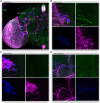
Glass-expressing cells contact both l-LNvs and s-LNvs. Immunostaining of GMR>transTANGO adult male brain. transTANGO marks presynaptic glass-expressing cells with GFP and the postsynaptic partners of those cells with RFP. Anti-PDF immunostaining confirms that both small and large LNvs are postsynaptic to GMR-Gal4 recruited cells. (A) Hemibrain. Asterisks were placed where PDF and RFP labeling co-localize: in posterior optic tract (POT) coming from l-LNvs, s-LNvs, and their terminals in the dorsal brain. (B) l-LNvs somatas (labeled with PDF, blue) have postsynaptic mark (RFP, magenta), HB eyelets projections can be seen nearby (labeled with GFP, green). (C,D) s-LNvs somatas and terminals have postsynaptic mark (RFP, magenta). Every brain analyzed (n = 9) showed similar staining. The images were acquired from different brains.
FIGURE 11
FIGURE 11
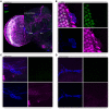
L2 interneurons form direct synaptic contacts with large but not small LNvs. Immunostaining of L2>transTANGO adult male brain. (A) Hemibrain. Asterisks mark places where PDF and RFP labeling co-localize, which means l-LNvs cell bodies and POT. (B) l-LNvs somatas have postsynaptic labeling, meaning that l-LNvs form direct contacts with L2 interneurons. This was seen in 86% of brains (n = 7). A L2 projection can be seen in cyan, rounding a somata. (C,D) Neither somatas nor projections of the s-LNvs have postsynaptic labeling. None of the brains analyzed showed RFP signal in these cells. The images were acquired from different brains.

Similar articles

Cited by

References

    1. Chatterjee A., Hardin P. E. (2010). Time to taste: circadian clock function in the Drosophila gustatory system. Fly 4 283–287. 10.4161/fly.4.4.13010 - DOI - PMC - PubMed
    1. Chatterjee A., Tanoue S., Houl J. H., Hardin P. E. (2010). Regulation of gustatory physiology and appetitive behavior by the Drosophila circadian clock. Curr. Biol. 20 300–309. 10.1016/j.cub.2009.12.055 - DOI - PMC - PubMed
    1. Chow E. S., Long D. M., Giebultowicz J. M. (2016). Circadian rhythm in mRNA expression of the glutathione synthesis gene Gclc is controlled by peripheral glial clocks in Drosophila melanogaster. Physiol. Entomol. 41 369–377. 10.1111/phen.12164 - DOI - PMC - PubMed
    1. Chung B. Y., Kilman V. L., Keath J. R., Pitman J. L., Allada R. (2009). The GABAA receptor RDL acts in peptidergic PDF neurons to promote sleep in Drosophila. Curr. Biol. 19 386–390. 10.1016/j.cub.2009.01.040 - DOI - PMC - PubMed
    1. Damulewicz M., Mazzotta G. M., Sartori E., Rosato E., Costa R., Pyza E. M. (2017). Cryptochrome is a regulator of synaptic plasticity in the visual system of Drosophila melanogaster. Front. Mol. Neurosci. 10:165. 10.3389/fnmol.2017.00165 - DOI - PMC - PubMed

LinkOut - more resources