Review
. 2020 Aug 4:11:1438.
doi: 10.3389/fimmu.2020.01438. eCollection 2020.

Role of Cystic Fibrosis Bronchial Epithelium in Neutrophil Chemotaxis

Affiliations
Review

Role of Cystic Fibrosis Bronchial Epithelium in Neutrophil Chemotaxis

Giulio Cabrini et al. Front Immunol. .

Abstract

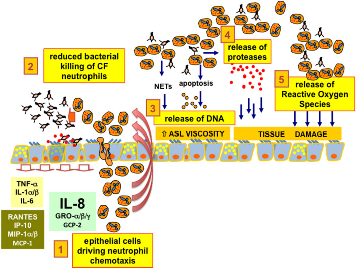
A hallmark of cystic fibrosis (CF) chronic respiratory disease is an extensive neutrophil infiltrate in the mucosa filling the bronchial lumen, starting early in life for CF infants. The genetic defect of the CF Transmembrane conductance Regulator (CFTR) ion channel promotes dehydration of the airway surface liquid, alters mucus properties, and decreases mucociliary clearance, favoring the onset of recurrent and, ultimately, chronic bacterial infection. Neutrophil infiltrates are unable to clear bacterial infection and, as an adverse effect, contribute to mucosal tissue damage by releasing proteases and reactive oxygen species. Moreover, the rapid cellular turnover of lumenal neutrophils releases nucleic acids that further alter the mucus viscosity. A prominent role in the recruitment of neutrophil in bronchial mucosa is played by CF bronchial epithelial cells carrying the defective CFTR protein and are exposed to whole bacteria and bacterial products, making pharmacological approaches to regulate the exaggerated neutrophil chemotaxis in CF a relevant therapeutic target. Here we revise: (a) the major receptors, kinases, and transcription factors leading to the expression, and release of neutrophil chemokines in bronchial epithelial cells; (b) the role of intracellular calcium homeostasis and, in particular, the calcium crosstalk between endoplasmic reticulum and mitochondria; (c) the epigenetic regulation of the key chemokines; (d) the role of mutant CFTR protein as a co-regulator of chemokines together with the host-pathogen interactions; and (e) different pharmacological strategies to regulate the expression of chemokines in CF bronchial epithelial cells through novel drug discovery and drug repurposing.

Keywords: chemotaxis; cystic fibrosis; epithelium; inflammation; lung; neutrophil.


Figures

Figure 1
Figure 1
CF bronchial epithelial cells and neutrophil chemotaxis. 1. epithelial cells driving neutrophil chemotaxis: bronchial epithelial cells drive the chemotaxis of neutrophils inside the lumen of bronchi and bronchioles of CF patients, mainly secreting the chemokine IL-8 (CXCL8) expressed upon interaction with bacteria with ASGM1R and TLR2/5 receptors exposed on the apical membrane. 2. reduced bacterial killing of CF neutrophils: CF neutrophils present different degrees of defective killing of the bacteria in the process of phagocytosis, being unable to clear completely the recurrent and, later on, chronic bacterial infection in the airways. 3. release of DNA: CF neutrophils are continuously stimulated in releasing Neutrophil Extracellular Traps. Despite the expected increase in efficiency of bacterial clearing, DNA from NETs increases the viscosity of the Airway Surface Liquid (ASL), further worsening the beat of cilia and the effectiveness of the CF mucociliary clearance. Moreover, CF neutrophils further release DNA as a consequence of apoptosis or necrosis due to the hypoxic environment resulting from mucus plugging of the conductive airways. 4. release of proteases: CF neutrophils exposed to bacteria or bacteria-derived components are prone to exocytosis of granules containing a wide armamentarium of proteases that are usually utilized by neutrophils to disrupt the tissues to allow their migration. In particular, an abundant amount of elastase has been found in CF lungs and its concentration has been related to CF lung disease progression and severity. 5. release of Reactive Oxygen Species: the unbalanced pro-oxidant milieu of the CF ASL due to excessive ROS derives mainly from “frustrated phagocytosis” of CF neutrophils and contributes together with proteases to a neutrophil-dependent airway tissue damage, a clear side effect dependent on the CFTR defect and worsened by an exaggerated neutrophil chemotaxis in CF bronchial lumen.
Figure 2
Figure 2
Selected examples of the miRNA network regulating IL-8. 1. microRNAs regulating the AKT-dependent NF-kB: miR-155-5p expression is potentiated by NF-kB and is elevated in CF lung epithelial cells and circulating CF neutrophils biopsied from CF patients. High levels of miR-155 specifically reduce the levels of SHIP1, thereby promoting PI3K/Akt activation. Phospho-Akt levels are therefore elevated in CF lung epithelial cells and can be specifically lowered by either antagomir-155 or elevated expression of SHIP1. Elevated miR-155-5p contributes to the pro-inflammatory expression of IL-8 in CF lung epithelial cells by lowering SHIP1 expression and thereby activating the PI3K/Akt signaling pathway (104). 2. transcriptional regulation of IL-8 gene expression: IL-8 promoter is under the control of transcription factors enhancing IL-8 mRNA production (examples are NF-κB, AP-1, C/EBPβ, CHOP, and CREB); furthermore, IL-8 gene expression is repressed by Oct-1 and by NF-κB repressing factor (NRF). All these transcription factors are regulated by microRNAs. 3. post-transcriptional miRNA-dependent regulation of IL-8: Fabbri et al. identified miR-93 as a miRNA that is decreased in cystic fibrosis IB3-1 and Cufi-1 cells infected with P. aeruginosa. The possible involvement of miR-93 in IL-8 gene regulation was validated using luciferase vectors and the results obtained indicate that IL-8 protein expression is post-transcriptionally regulated by interactions of the IL-8 mRNA with the inhibitory miR-93 (105). The involvement of microRNAs in IL-8 is also supported by the study of Oglesby et al. (106) who identified miR-17 as a miRNA regulating IL-8. Interestingly, its expression was decreased in adult CF bronchial brushings and bronchial epithelial cells chronically stimulated with Pseudomonas-conditioned medium (106). 5′-UTR, 3′-UTR: 5′ and 3′ untranslated region of the IL-8 mRNA; CDS, coding sequences.

References

    1. Riordan JR, Rommens JM, Kerem B, Alon N, Rozmahel R, Grzelczak Z, et al. Identification of the cystic fibrosis gene: cloning and characterization of complementary DNA. Science. (1989) 245:1066–73. 10.1126/science.2475911 - DOI - PubMed
    1. Rommens JM, Iannuzzi MC, Kerem B, Drumm ML, Melmer G, Dean M, et al. Identification of the cystic fibrosis gene: chromosome walking and jumping. Science. (1989) 245:1059–65. 10.1126/science.2772657 - DOI - PubMed
    1. Kerem B, Rommens JM, Buchanan JA, Markiewicz D, Cox TK, Chakravarti A, et al. Identification of the cystic fibrosis gene: genetic analysis. Science. (1989) 245:1073–80. 10.1126/science.2570460 - DOI - PubMed
    1. Khan TZ, Wagener JS, Bost T, Martinez J, Accurso FJ, Riches DW. Early pulmonary inflammation in infants with cystic fibrosis. Am J Respir Crit Care Med. (1995) 151:1075–82. 10.1164/ajrccm.151.4.7697234 - DOI - PubMed
    1. Noah TL, Black HR, Cheng PW, Wood RE, Leigh MW. Nasal and bronchoalveolar lavage fluid cytokines in early cystic fibrosis. J Infect Dis. (1997) 175:638–47. 10.1093/infdis/175.3.638 - DOI - PubMed

Publication types

Substances