Observational Study
. 2020 Aug 11:11:1765.
doi: 10.3389/fimmu.2020.01765. eCollection 2020.

Eosinophilia Associated With CD3-CD4+ T Cells: Characterization and Outcome of a Single-Center Cohort of 26 Patients

Affiliations
Observational Study

Eosinophilia Associated With CD3-CD4+ T Cells: Characterization and Outcome of a Single-Center Cohort of 26 Patients

Caroline Carpentier et al. Front Immunol. .

Abstract

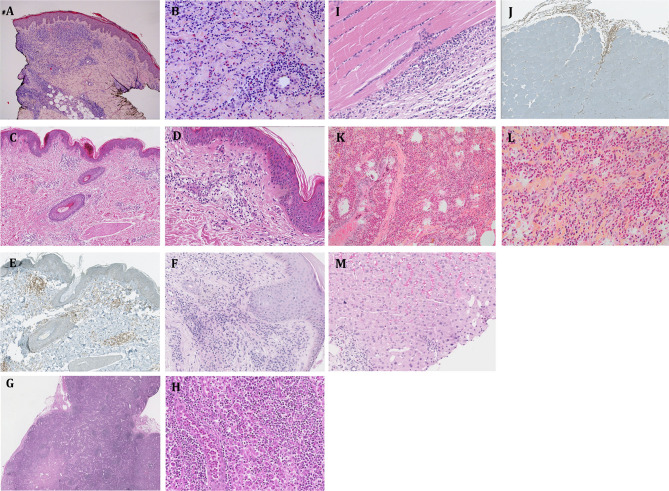
Background: Lymphocytic variant hypereosinophilic syndrome is characterized by marked over-production of eosinophilopoietic factor(s) by dysregulated T cells leading to eosinophil expansion. In most cases, these T cells are clonal and express a CD3-CD4+ phenotype. As this is a rare disorder, presenting manifestations, disease course, treatment responses, and outcome are not well-characterized. Materials and Methods: In this retrospective single-center observational study, we reviewed medical files of all patients with persistent hypereosinophilia seen between 1994 and 2019 in whom CD3-CD4+ T cells were detected. Data collection included clinical and biological findings at presentation, treatment responses, disease course, and serial CD3-CD4+ T cell counts. Results: Our cohort comprises 26 patients, including 2 with hypereosinophilia of undetermined significance. All 24 symptomatic patients had cutaneous lesions and/or angioedema, and fasciitis was present in several cases. The aberrant T cell subset represented 2% or less total lymphocytes in 11 subjects. TCR gene rearrangement patterns on whole blood were polyclonal in these cases, while they all had serum CCL17/TARC levels above 1,500 pg/ml. Disease manifestations were mild and did not require maintenance therapy in roughly one third of the cohort, while two thirds required long-term oral corticosteroids and/or second-line agents. Among these, interferon-alpha was the most effective treatment option with a response observed in 8/8 patients, one of whom was cured of disease. Treatment had to be interrupted in most cases however due to poor tolerance and/or development of secondary resistance. Anti-interleukin-5 antibodies reduced blood eosinophilia in 5/5 patients, but clinical responses were disappointing. A sub-group of 5 patients had severe treatment-refractory disease, and experienced significant disease- and treatment-related morbidity and mortality, including progression to T cell lymphoma in three. Conclusions: This retrospective longitudinal analysis of the largest monocentric cohort of CD3-CD4+ T cell associated lymphocytic variant hypereosinophilic syndrome published so far provides clinicians confronted with this rare disorder with relevant new data on patient presentation and outcome that should help tailor therapy and follow-up to different levels of disease severity. It highlights the need for novel therapeutic options, especially for the subset of patients with severe treatment-refractory disease. Future research efforts should be made toward understanding CD3-CD4+ T cell biology in order to develop new treatments that target primary pathogenic mechanisms.

Keywords: CD3−CD4+ T cells; T cell lymphoma; TARC (CCL17); anti-interleukin-5; fasciitis; interferon-alpha; interleukin-5; lymphocytic variant hypereosinophilic syndrome.


Figures

Figure 1
Figure 1
Spectrum of cutaneous and soft tissue manifestations observed in patients with CD3CD4+ T cell associated L-HES. (A) P12, rash on arm lasting less than 1 day. (B) P13, confluent localized papules on abdomen lasting several days. (C,D) P3, disseminated migratory papules on back and arm. (E) P3, large fixed cutaneous plaque lesion on lower leg lasting months. (F) P16, urticaria on thigh, lasting minutes. (G) P16, two fixed pigmented plaques around nipple. (H) P21, fixed plaque. (I) P25, disseminated migratory papules on abdomen. (J) P2, scaly eczema hands lasting years. (K) P10, feet between and during flares of episodic angioedema. (L) P14, diffuse angioedema of lower leg with more localized lumps lasting several hours. (M) P21, diffusely lichenified hyper-pigmented skin.
Figure 2
Figure 2
Non-cutaneous complications of CD3CD4+ T cell associated L-HES. (A) P3, arthritis and synovitis right ankle. (B) P11, fasciitis—MRI showing gadolinium enhancement of the fascia and underlying sartorius and gracilis muscles of right leg. (C,D) P13, pulmonary involvement—Thoracic CT (C) scan showing nodule in left lower lobe (see white arrow), with mild to moderate hypermetabolic activity shown by FDG-PET scan (D).
Figure 3
Figure 3
Biopsy findings in patients with CD3CD4+ T cell-associated L-HES. (A,B) P3, skin biopsy (thigh) at site of persistent papular eruption despite OCS therapy: hematoxylin-eosin stain showing dense lymphocytic and eosinophilic infiltrate of upper and lower dermis (blood eosinophils elevated); 25-fold (A) and 200-fold (B) magnification. (C–E) P25, skin biopsy (thigh) at site of papular eruption before initiation of OCS therapy: hematoxylin-eosin stain showing moderate lymphocytic infiltrate of upper and lower dermis in the absence of eosinophils (although marked blood hypereosinophilia present at the time); combined immuno-staining for CD4 (brown) and CD8 (red) (E) showing that the lymphocytes are CD4 cells; 40-fold (C,E) and 100-fold (D) magnification. (F) P6, skin biopsy (popliteal region) at site of persistent fixed eczematous plaque despite ongoing OCS treatment: hematoxylin-eosin stain showing a dense lymphocytic infiltrate of the upper and lower dermis (blood eosinophils normal); 100-fold magnification. (G, H) P3, biopsy of enlarged lymph node (inguinal region): hematoxylin-eosin stain showing a dense inflammatory infiltrate and marked dilatation of sinusoid vessels containing numerous eosinophils; 25-fold (G) and 200-fold (H) magnification (I,J) P25, fascia-muscle biopsy (thigh): hematoxylin-eosin stain showing a dense inflammatory infiltrate of the fascia and perimysium, composed of lymphocytes, eosinophils, and plasma cells; combined immuno-staining for CD4 (brown) and CD8 (red) (J) showing that the lymphocytes are CD4 cells; 200-fold (I) and 40-fold (J) magnification. (K,L) P13, lung biopsy: hematoxylin-eosin-safran stain showing a dense intra-alveolar eosinophilic infiltrate; 100-fold (K) and 200-fold (L) magnification. (M) P17, liver biopsy: hematoxylin-eosin stain showing an intrasinusoidal and portal-space inflammatory infiltrate composed of lymphocytes and eosinophils; 200-fold magnification.
Figure 4
Figure 4
T cell phenotyping by flow cytometry in 2 representative patients with a CD3CD4+ T cell subset. (A) T cell phenotyping by flow cytometry showing a large (P12, top) and a small (P19, bottom) CD3-CD4+ T cell subset. The plots show the gating strategy from left to right: gate on lymphocytes on the basis of forward- and side-scatter, after selecting CD45hi cells (not shown) CD3 expression is plotted against side-scatter and CD3+ cells (green) are differentiated from CD3- cells (red), the proportions of CD3+CD4+ and CD3CD4+ T cells among CD45hi cells are shown on the right. (B) Intracytoplasmic IL-5 expression was assessed in CD4 T cells from patient P19. The histogram overlay shows IL-5 expression in CD3+CD4+ T cells (green) and in CD3CD4+ cells (red).
Figure 5
Figure 5
Treatment regimens and responses in patients with CD3CD4+ T cell associated L-HES. (A) Proportions of patients who required no maintenance treatment (white), prolonged OCS monotherapy (gray), or addition of a second-line agent (blue) to control disease. (B) shows the minimally effective dose of oral CS as monotherapy for a complete response early in disease course in the 13 patients for whom this dose could be determined accurately (one patient was already treated with HU when OCS were initiated, and OCS could not be increased to a dose such that a complete response occurred in two others due to marked intolerance). Proportions of patients who experienced a complete response with a PDN-equivalent dose at 10 mg or less (light gray), 11–20 mg (medium gray), and above 20 mg (dark gray) daily are depicted. (C) shows responses to the most commonly administered second-line agents, separating biological (left) from clinical (right) responses. A partial response is defined as a reduction of eosinophil counts by at least 50% with treatment (biological) and persistent but improved symptoms (clinical). A complete response is defined as normalization of blood eosinophil counts (biological) and resolution of HES-related symptoms (clinical). CSA cyclosporin A, HU hydroxyurea, IFN interferon, UNK unknown. See Supplementary Table 5 for individual data.
Figure 6
Figure 6
Evolution of absolute CD3CD4+ T cell counts over time. Evolution of absolute blood CD3CD4+ T cell counts over time is shown for all patients except P22 and P23 for whom duration of follow-up is less than a year, and P24 who is followed in another center. Absolute CD3CD4+ T cell counts are normalized relative to baseline counts for each patient (i.e., those at initial detection of these cells), and are shown separately for untreated subjects (A) and subjects receiving treatment (B) either with oral CS alone (blue lines) or second-line therapy (yellow lines). For the three patients who developed lymphoma (P1, P2, P10) the count at last follow-up is that at the time lymphoma was diagnosed. For the other patients, the most recent CD3CD4+ cell count and percentage, as well as the interval since first detection, are shown in Supplementary Table 7. Patients who were treated at baseline are indicated with an asterisk.

References

    1. Hardy WR, Anderson RE. The hypereosinophilic syndromes. Ann Intern Med. (1968) 68:1220–9. 10.7326/0003-4819-68-6-1220 - DOI - PubMed
    1. Chusid MJ, Dale DC, West BC, Wolff SM. The hypereosinophilic syndrome: analysis of fourteen cases with review of the literature. Medicine. (1975) 54:1–27. 10.1097/00005792-197501000-00001 - DOI - PubMed
    1. Valent P, Klion AD, Horny HP, Roufosse F, Gotlib J, Weller PF, et al. . Contemporary consensus proposal on criteria and classification of eosinophilic disorders and related syndromes. J Allergy Clin Immunol. (2012) 130:607–12.e9. 10.1016/j.jaci.2012.02.019 - DOI - PMC - PubMed
    1. Cogan E, Schandene L, Crusiaux A, Cochaux P, Velu T, Goldman M. Brief report: clonal proliferation of type 2 helper T cells in a man with the hypereosinophilic syndrome. N Engl J Med. (1994) 330:535–8. 10.1056/NEJM199402243300804 - DOI - PubMed
    1. Lefevre G, Copin MC, Staumont-Salle D, Avenel-Audran M, Aubert H, Taieb A, et al. . The lymphoid variant of hypereosinophilic syndrome: study of 21 patients with CD3-CD4+ aberrant T-cell phenotype. Medicine. (2014) 93:255–66. 10.1097/MD.0000000000000088 - DOI - PMC - PubMed

Publication types