Review
. 2020 Aug 7:11:1949.
doi: 10.3389/fimmu.2020.01949. eCollection 2020.

Overview of Immune Response During SARS-CoV-2 Infection: Lessons From the Past

Affiliations
Review

Overview of Immune Response During SARS-CoV-2 Infection: Lessons From the Past

Vibhuti Kumar Shah et al. Front Immunol. .

Abstract

After the 1918 flu pandemic, the world is again facing a similar situation. However, the advancement in medical science has made it possible to identify that the novel infectious agent is from the coronavirus family. Rapid genome sequencing by various groups helped in identifying the structure and function of the virus, its immunogenicity in diverse populations, and potential preventive measures. Coronavirus attacks the respiratory system, causing pneumonia and lymphopenia in infected individuals. Viral components like spike and nucleocapsid proteins trigger an immune response in the host to eliminate the virus. These viral antigens can be either recognized by the B cells or presented by MHC complexes to the T cells, resulting in antibody production, increased cytokine secretion, and cytolytic activity in the acute phase of infection. Genetic polymorphism in MHC enables it to present some of the T cell epitopes very well over the other MHC alleles. The association of MHC alleles and its downregulated expression has been correlated with disease severity against influenza and coronaviruses. Studies have reported that infected individuals can, after recovery, induce strong protective responses by generating a memory T-cell pool against SARS-CoV and MERS-CoV. These memory T cells were not persistent in the long term and, upon reactivation, caused local damage due to cross-reactivity. So far, the reports suggest that SARS-CoV-2, which is highly contagious, shows related symptoms in three different stages and develops an exhaustive T-cell pool at higher loads of viral infection. As there are no specific treatments available for this novel coronavirus, numerous small molecular drugs that are being used for the treatment of diseases like SARS, MERS, HIV, ebola, malaria, and tuberculosis are being given to COVID-19 patients, and clinical trials for many such drugs have already begun. A classical immunotherapy of convalescent plasma transfusion from recovered patients has also been initiated for the neutralization of viremia in terminally ill COVID-19 patients. Due to the limitations of plasma transfusion, researchers are now focusing on developing neutralizing antibodies against virus particles along with immuno-modulation of cytokines like IL-6, Type I interferons (IFNs), and TNF-α that could help in combating the infection. This review highlights the similarities of the coronaviruses that caused SARS and MERS to the novel SARS-CoV-2 in relation to their pathogenicity and immunogenicity and also focuses on various treatment strategies that could be employed for curing COVID-19.

Keywords: COVID-19; HLA; MHC presentation; T cells; coronavirus; immune response; memory T cell.


Figures

Figure 1
Figure 1
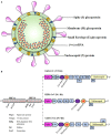
Schematic representation of the coronavirus structure and genomic comparison of coronaviruses. (A) Representation of coronavirus showing different components of the particle, which is 100–160 nm in diameter. The single-stranded RNA (ssRNA) genome, covered with the envelope and membrane proteins, gains access into the host cell and hijacks the replication machinery. (B) The ssRNA of SARS-CoV-2 is about 30 kb and has similarities with the genomes of SARS-CoV and MERS-CoV. Translation of this ssRNA results in the formation of two polyproteins, namely pp1a and pp1ab, that are further sliced to generate numerous non-structural proteins (NSPs). The remaining ORFs encode for various structural and accessory proteins that help in assembly of the viral particle and evading immune response.
Figure 2
Figure 2
Plausible host immune responses during COVID-19 infection. The SARS-CoV-2 virus infects through the naso-oral route, followed by infection in cells expressing ACE2 receptor in the lung, such as type 2 alveolar cells. These viruses dampen anti-viral IFN responses by evading the innate immune cells as a consequence of unrestrained virus replication. The infiltration of monocytes/macrophages, neutrophils, and several other adaptive immune cells leads to increased pro-inflammatory cytokines. In the helper T cell subset, stimulation of Th1/Th17 cells with viral epitopes may lead to aggravated inflammatory responses. This inflammatory response results in “cytokine storms” that lead to immunopathologies like pulmonary edema and pneumonia. Cytotoxic T cells recruited to the site of infection try to kill virus-infected cells in the lungs. B cells/plasma cells also recognize viral proteins and are activated to produce antibodies specific to SARS-CoV-2, which may help in deactivating viruses and provide systemic immunity in different organs.
Figure 3
Figure 3
SARS-CoV-mediated evasion of host innate immune response. The viral antigens are recognized via different PRRs to elicit the innate immune response. (1) Upon interaction of the virus with surface PRRs or the specific receptor, the particles are endocytosed into the cytosol and are then recognized by cytosolic PRRs like RIG-I and MDA5. (2) The viral genome, along with different proteins, interacts with MAVS and initiates NF-κB activation via triggering a signaling cascade that involves numerous E3 ubiquitin kinases and ligases. (3) Upon translocation into the nucleus, activated NF-κB acts as a transcriptional activator for numerous pro-inflammatory cytokines with an NF-κB-response element. The IFN-regulatory factor 3 (IRF3), upon phosphorylation via ubiquitin kinases, homodimerizes and moves inside the nucleus to activate the transcription of Type I IFNs. (4) Type I IFNs have both autocrine and paracrine mechanisms to activate the JAK–STAT signaling pathway via IFNα/β receptor (IFNAR), followed by phosphorylation of STAT1 and STAT2 via cytoplasmic protein JAK1 and TYK2 kinases. STAT1 & STAT2 heterodimers translocate into the nucleus and are recruited for transcription of the IFN-stimulated gene having an IFN-stimulated response element (ISRE) present on their promoter. SARS-CoV and other coronaviruses have found many ways to inhibit the signaling cascade by utilizing either the structural proteins (M and N protein) or NSPs (NSP1, NSP3b, and NSP6 along with PLpro), shown as numbers and letters in the figure. Together, the production of pro-inflammatory cytokines and type I IFNs tries to create an antiviral immune microenvironment that controls viral synthesis and infection, but the viruses have deployed various strategies to shut down these signaling pathways to counteract the immune response. RIG-I, Retinoic acid-Inducible Gene I protein; MDA5, Melanoma Differentiation-Associated protein 5; MAVS, Mitochondrial antiviral-signaling protein; M, Membrane protein; N, Nucleocapsid; IFNAR, IFNα/β receptor; ISGs, IFN-stimulated genes; ISRE, IFN-stimulated response element.

References

    1. Zhou P, Yang X-L, Wang XG, Hu B, Zhang L, Zhang W, et al. . A pneumonia outbreak associated with a new coronavirus of probable bat origin. Nature. (2020) 579:270–3. 10.1038/s41586-020-2012-7 - DOI - PMC - PubMed
    1. Lu R, Zhao X, Li J, Niu P, Yang B, Wu H, et al. . Genomic characterisation and epidemiology of 2019 novel coronavirus: implications for virus origins and receptor binding. Lancet. (2020) 395:565–74. 10.1016/S0140-6736(20)30251-8 - DOI - PMC - PubMed
    1. Ong SWX, Tan YK, Chia PY, Lee TH, Ng OT, Wong MSY, et al. . Air, surface environmental, and personal protective equipment contamination by severe acute respiratory syndrome coronavirus 2 (SARS-CoV-2) from a symptomatic patient. JAMA - J Am Med Assoc. (2020) 323:5–7. 10.1001/jama.2020.3227 - DOI - PMC - PubMed
    1. de Wit E, van Doremalen N, Falzarano D, Munster VJ. SARS and MERS : recent insights into emerging coronaviruses. Nat Rev Microbiol. (2016) 14:523–34. 10.1038/nrmicro.2016.81 - DOI - PMC - PubMed
    1. Verity R, Okell LC, Dorigatti I, Winskill P, Whittaker C, Imai N, et al. . Estimates of the severity of coronavirus disease 2019 : a model-based analysis. Lancet Infect Dis. (2020) 3099:1–9. 10.1016/S1473-3099(20)30243-7 - DOI - PMC - PubMed

Publication types

MeSH terms