

Pseudogene DUXAP8 Promotes Cell Proliferation and Migration of Hepatocellular Carcinoma by Sponging MiR-490-5p to Induce BUB1 Expression
- PMID: 32849765
- PMCID: PMC7396656
- DOI: 10.3389/fgene.2020.00666
Pseudogene DUXAP8 Promotes Cell Proliferation and Migration of Hepatocellular Carcinoma by Sponging MiR-490-5p to Induce BUB1 Expression
Abstract
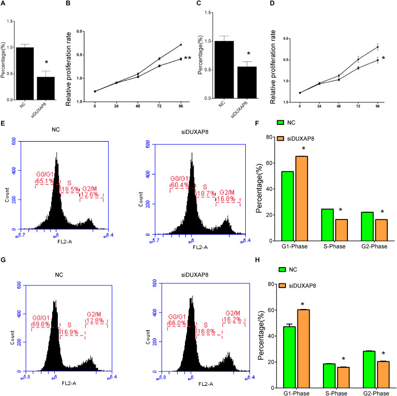
Hepatocellular Carcinoma (HCC) currently remains one of the most lethal malignancies worldwide. Recently, long non-coding RNAs (lncRNAs) had been demonstrated to play a crucial role in the progression of multiple human cancers, including HCC. In this study, we found that lncRNA DUXAP8 was upregulated in tumor samples and served as an oncogene in HCC. Bioinformatics analysis showed that DUXAP8 was significantly associated with the regulation of centrosome organization, homologous recombination, meiotic cell cycle process, sister chromatid segregation, nuclear chromosome segregation, and RNA export from the nucleus. The knockdown of DUXAP8 significantly suppresses cell proliferation and the cell cycle but induces cell apoptosis in HCC. Mechanically, the present study showed that DUXAP8 serves as a sponge of MiR-490-5p to promote the expression of BUB1 in HCC. Although the underlying regulatory mechanisms of DUXAP8 in HCC require further investigation, this study, for the first time, showed that DUXAP8 can serve as a new therapeutic target for HCC.
Keywords: Hepatocellular Carcinoma; bioinformatics; long non-coding RNA; regulatory mechanism; target.
Copyright © 2020 Zhang, Chu, Zheng, Ren and Tian.
Figures


Similar articles
-
Long Non-coding RNA Double Homeobox A Pseudogene 8: A Novel Oncogenic Propellant in Human Cancer.Front Cell Dev Biol. 2021 Sep 23;9:709069. doi: 10.3389/fcell.2021.709069. eCollection 2021. Front Cell Dev Biol. 2021. PMID: 34631702 Free PMC article. Review.
-
Long non-coding RNA DUXAP8 promotes tumorigenesis by regulating IGF1R via miR-9-3p in hepatocellular carcinoma.Exp Ther Med. 2021 Jul;22(1):755. doi: 10.3892/etm.2021.10187. Epub 2021 May 12. Exp Ther Med. 2021. PMID: 34035852 Free PMC article.
-
m6A Modification-Mediated DUXAP8 Regulation of Malignant Phenotype and Chemotherapy Resistance of Hepatocellular Carcinoma Through miR-584-5p/MAPK1/ERK Pathway Axis.Front Cell Dev Biol. 2021 Dec 9;9:783385. doi: 10.3389/fcell.2021.783385. eCollection 2021. Front Cell Dev Biol. 2021. PMID: 34957112 Free PMC article.
-
lncRNA DUXAP8 Facilitates Multiple Malignant Phenotypes and Resistance to PARP Inhibitor in HCC via Upregulating FOXM1.Mol Ther Oncolytics. 2020 Oct 22;19:308-322. doi: 10.1016/j.omto.2020.10.010. eCollection 2020 Dec 16. Mol Ther Oncolytics. 2020. PMID: 33313387 Free PMC article.
-
Clinical significance of long non-coding RNA DUXAP8 and its protein coding genes in hepatocellular carcinoma.J Cancer. 2020 Aug 25;11(20):6140-6156. doi: 10.7150/jca.47902. eCollection 2020. J Cancer. 2020. PMID: 32922554 Free PMC article.
Cited by
-
LncRNA DUXAP8 as a prognostic biomarker for various cancers: A meta-analysis and bioinformatics analysis.Front Genet. 2022 Aug 15;13:907774. doi: 10.3389/fgene.2022.907774. eCollection 2022. Front Genet. 2022. PMID: 36046244 Free PMC article.
-
Long Non-coding RNA Double Homeobox A Pseudogene 8: A Novel Oncogenic Propellant in Human Cancer.Front Cell Dev Biol. 2021 Sep 23;9:709069. doi: 10.3389/fcell.2021.709069. eCollection 2021. Front Cell Dev Biol. 2021. PMID: 34631702 Free PMC article. Review.
-
The World of Pseudogenes: New Diagnostic and Therapeutic Targets in Cancers or Still Mystery Molecules?Life (Basel). 2021 Dec 7;11(12):1354. doi: 10.3390/life11121354. Life (Basel). 2021. PMID: 34947885 Free PMC article. Review.
-
Development and validation of ferroptosis-related lncRNAs prognosis signatures in kidney renal clear cell carcinoma.Cancer Cell Int. 2021 Nov 4;21(1):591. doi: 10.1186/s12935-021-02284-1. Cancer Cell Int. 2021. PMID: 34736453 Free PMC article.
-
Functions of lncRNA DUXAP8 in non-small cell lung cancer.Mol Biol Rep. 2022 Mar;49(3):2531-2542. doi: 10.1007/s11033-021-07066-6. Epub 2022 Jan 15. Mol Biol Rep. 2022. PMID: 35031926 Review.
References
-
- Lettnin A. P., Wagner E. F., Carrett-Dias M., Dos Santos Machado K., Werhli A., Canedo A. D., et al. (2019). Silencing the OCT4-PG1 pseudogene reduces OCT-4 protein levels and changes characteristics of the multidrug resistance phenotype in chronic myeloid leukemia. Mol. Biol. Rep. 46 1873–1884. 10.1007/s11033-019-04639-4 - DOI - PubMed
LinkOut - more resources
Full Text Sources