. 2020 Aug 11:2020:9108317.
doi: 10.1155/2020/9108317. eCollection 2020.

Comparison of Different Types of Corneal Foreign Bodies Using Anterior Segment Optical Coherence Tomography: A Prospective Observational Study

Affiliations

Comparison of Different Types of Corneal Foreign Bodies Using Anterior Segment Optical Coherence Tomography: A Prospective Observational Study

Tao Wang et al. J Ophthalmol. .

Abstract

Purpose: The present study highlighted the value of anterior segment optical coherence tomography (AS-OCT) for different types of corneal foreign bodies in humans.

Methods: This study was a prospective observational study. The patients included were divided into two groups. If the patients were directly diagnosed based on eye injury history and slit-lamp examination, then they were assigned to Group A. Otherwise, the patients were assigned to Group B. We compared and described the characteristics of the corneal foreign body in both groups using AS-OCT.

Results: From October 2017 to January 2020, 36 eyes of 36 patients (9 females and 27 males) with a mean age of 37.8 ± 11.7 years were included in the study. Patients in Group A were the majority and accounted for 72.2% (26/36). High signals on AS-OCT images were the main constituent and accounted for 92.3% (24/26) in Group A and 70.0% (7/10) in Group B. Most of the patients in Group A, 96.2% (25/26), had clear boundaries. A blurred boundary was observed in 70.0% (7/10) of the patients in Group B. The foreign bodies on AS-OCT images had key characteristics of a high signal followed by a central zone shadowing effect and a low signal followed by a marginal zone shadowing effect. Further, all of the lesions could be directly located in Group B, and 92.3% (24/26) of the patients in Group A did not have directly located lesions. Six representative cases are described in detail.

Conclusions: AS-OCT is a valuable tool in the diagnosis and management of corneal foreign bodies, especially for unusual corneal foreign body.


Conflict of interest statement

The authors declare that they have no conflicts of interest.

Figures

Figure 1
Figure 1
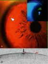
Anterior segment and AS-OCT image for Case 1. (a) Anterior segment photograph showed a corneal FB and inflammatory infiltration around the FB at an approximately 3:00 o'clock location approaching the limbus (arrowhead). (b) AS-OCT image showed that the FB showed a single high signal with clear boundary (arrow) following a central zone shadowing effect (star). The depth of the FB was estimated indirectly by subtracting the thickness of the underside of the high-reflected signal surrounding a FB (359 m) from the thickness of the cornea (662 m), which was less than 303 μm.
Figure 2
Figure 2
Anterior segment and AS-OCT image for Case 2. (a) Anterior segment photograph showing multiple small granular corneal lesions in the shallow cornea (arrowheads). (b) AS-OCT showed that there were three high signals with blurred boundary (arrows). Two of the signals had a central zone shadowing effect (stars). They were directly located and were 72, 86, and 114 μm beneath the corneal surface.
Figure 3
Figure 3
Anterior segment and AS-OCT image for Case 3. (a) Anterior segment photograph: at 9:00 o'clock inside the limbus (arrowhead), a light brown mass was observed without surface fluorescence staining, and fluorescein gathered around the edges. Neovascularization was also seen. (b) AS-OCT image showing that a crescent-shaped low reflective signal (arrowhead) with bilateral marginal zone shadowing (stars) was found 21 μm below the epithelium surface.
Figure 4
Figure 4
Anterior segment and AS-OCT image for Case 4. (a) Anterior segment photograph showing multiple cream-like corneal opacities (arrowheads) surrounding a wrinkled transparent cornea. (b) AS-OCT image showed multiple spines (high signals) with blurred boundary (arrows) on the superficial epithelium followed by a central zone shadowing effect (stars). The corneal thickness was 546 μm, within the normal range.
Figure 5
Figure 5
Anterior segment and AS-OCT image for Case 5. (a) Anterior segment photograph showing a patchy cloud-like haze over the cornea and a cloudy milky lump in front of the pupil (arrowhead). (b) AS-OCT scanning showed there was a lesion of high signal with blurred boundary (arrow) followed by a central zone shadowing effect (star). The lesion had a diameter of 1372 μm. The corneal thickness was 604 μm, and the epithelial thickness was 102 μm. A layer of continuous epithelium tissue was seen on the bottom of the lesion.
Figure 6
Figure 6
Anterior segment and AS-OCT image for Case 6. (a) Anterior segment photograph showed a slight bulge at 10:00 o'clock in the peripheral cornea (white arrowhead). Corneal fluorescein staining was negative (arrowhead). (b) AS-OCT scanning showed there was a lesion of high signal with blurred boundary (arrow) without a shadowing effect. The lesion was directly located and was 136 μm beneath the corneal surface.

Similar articles

Cited by

References

    1. McGwin G., Jr., Xie A., Owsley C. Rate of eye injury in the United States. Archives of Ophthalmology. 2005;123(7):970–976. doi: 10.1001/archopht.123.7.970. - DOI - PubMed
    1. McGwin G., Jr., Owsley C. Incidence of emergency department-treated eye injury in the United States. Archives of Ophthalmology. 2005;123(5):662–666. doi: 10.1001/archopht.123.5.662. - DOI - PubMed
    1. Guier C. P., Stokkermans T. J. StatPearls. Treasure Island, FL, USA: Treasure Island (FL): StatPearls Publishing; 2020. Cornea foreign body removal. - PubMed
    1. Girgis R., Verma S. Unusual corneal foreign body. Eye. 2009;23(7):p. 1609. doi: 10.1038/eye.2008.248. - DOI - PubMed
    1. Al-Ghadeer H. A., Al-Assiri A. Identification and localization of multiple intrastromal foreign bodies with anterior segment optical coherence tomography and ocular pentacam. International Ophthalmology. 2014;34(2):355–358. doi: 10.1007/s10792-013-9800-0. - DOI - PubMed