Case Reports
. 2020 Aug 8:2020:9056209.
doi: 10.1155/2020/9056209. eCollection 2020.

Metastatic Syringocystadenocarcinoma Papilliferum: A Case Report, Tumor Genomic Profiling, and Literature Review

Affiliations
Case Reports

Metastatic Syringocystadenocarcinoma Papilliferum: A Case Report, Tumor Genomic Profiling, and Literature Review

Erdem Altunel et al. Case Rep Oncol Med. .

Abstract

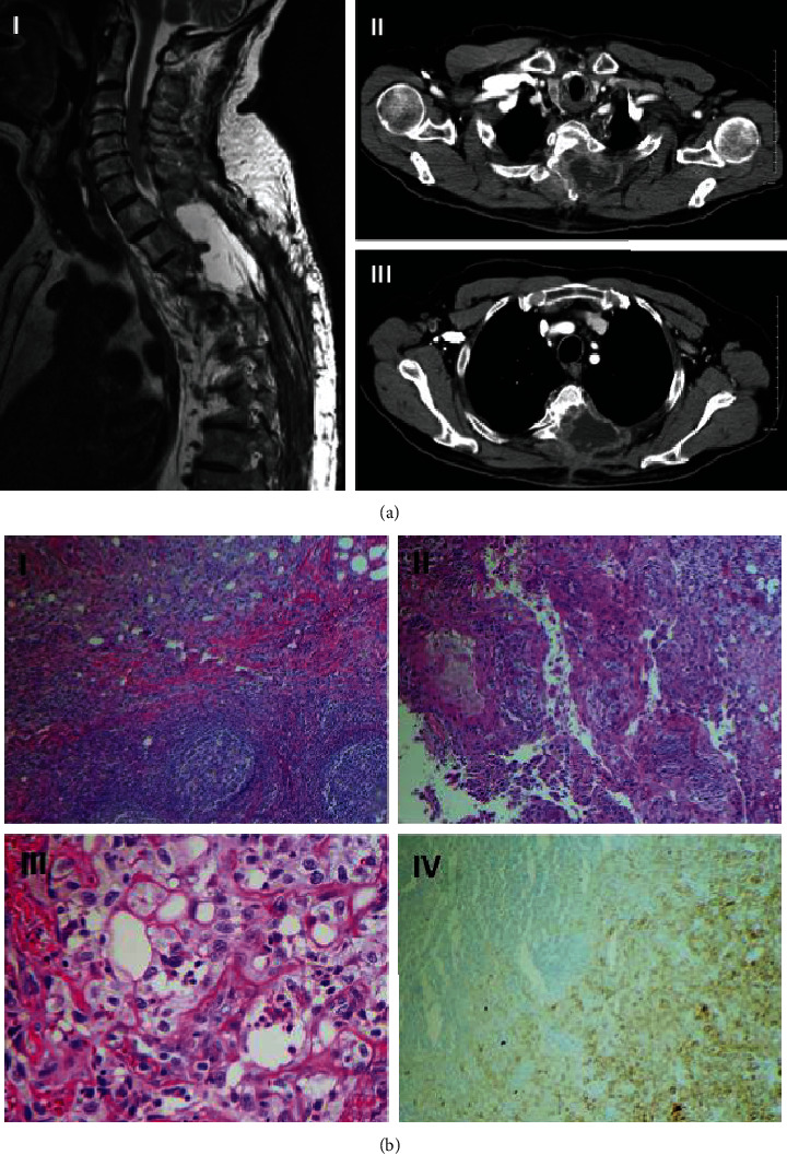
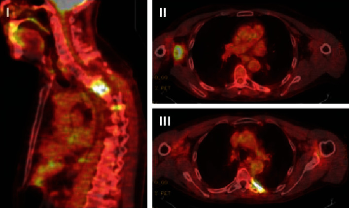
Syringocystadenocarcinoma papilliferum (SCACP) is an extremely rare cutaneous neoplasm of the apocrine or eccrine sweat glands. Solid and cystic glandular structures with cribriform and tubular architecture along with CK5/6, pankeratin and p63 immuno-profile set apart SCACP from other cutaneous malignancies. Wide local excision (WLE) has been the mainstay treatment for localized SCACP; however, no standard treatment has yet been established for unresectable or metastatic disease. Herein, we report a 74-year-old male with SCACP, who initially presented with a painful nodule on the upper back and later developed metastatic disease. He was treated with carboplatin and paclitaxel with concurrent intensity-modulated radiation therapy (IMRT), which resulted in disease stabilization for 12 months. Next generation sequencing (NGS) revealed a total of 18 genomic alterations associated with potential benefit from targeted therapeutics. PD-L1 expression was identified in 70% of tumor cells. These findings suggest that the opportunity of targeted therapeutics and immunotherapy exist as for metastatic SCACP. Reporting molecular profile of the rare tumors with no established standard treatment options should be encouraged.


Conflict of interest statement

The authors declare that they have no conflicts of interest.

Figures

Figure 1
Figure 1
Radiologic and histologic appearance of syringocystadenocarcinoma papilliferum. (a) (I) Magnetic resonance imaging showing soft tissue mass in the thoracic spine. (II) Axial computed tomography (CT) scan showing tumor invasion into the vertebrae at T4 level. (III) Axial CT scan showing tumor invasion into the vertebrae at T2 level and enlarged right axillary lymph node. (b) (I) Lymph node with metastatic tumor, H&E stain, 40x. (II) Solid and glandular structures with warty architecture, lined by poorly-differentiated epithelium, H&E stain, 100x. (III) Clusters and nests of highly pleomorphic cells with bizarre irregular nuclei and permanent nucleoli, entrapped in fibrotic stroma, consistent with carcinoma, H&E stain, 400x. (IV) Metastatic carcinoma cells are highlighted by immune-stain (right lower corner); residual uninvolved lymph node shows no immunopositivity (left upper corner), wide spectrum cytokeratin immunostain, 100x.
Figure 2
Figure 2
Positron emission tomography (PET) scan images 12 months after chemoradiotherapy. Hypermetabolic activity in the left paraspinal region at the level of T4-T5 (I), in the right axilla (II), and in the left sixth rib medially (III).

References

    1. Dissanayake R. V. P., Salm R. Sweat-gland carcinomas: prognosis related to histological type. Histopathology. 1980;4(4):445–466. doi: 10.1111/j.1365-2559.1980.tb02939.x. - DOI - PubMed
    1. Parekh V., Guerrero C. E., Knapp C. F., Elmets C. A., McKay K. M. A Histological Snapshot of Hypothetical Multistep Progression From Nevus Sebaceus to Invasive Syringocystadenocarcinoma Papilliferum. The American Journal of Dermatopathology. 2016;38(1):56–62. doi: 10.1097/DAD.0000000000000370. - DOI - PubMed
    1. Iga N., Fujii H., Miyake T., Ehara M., Kore-eda S. Syringocystadenocarcinoma Papilliferum in the perianal area. Case Reports in Dermatology. 2015;7(2):84–89. doi: 10.1159/000381940. - DOI - PMC - PubMed
    1. Zhang Y., Kong Y. Y., Cai X., Shen X. X., Kong J. C. Syringocystadenocarcinoma papilliferum: Clinicopathologic analysis of 10 cases. Journal of Cutaneous Pathology. 2017;44(6):538–543. doi: 10.1111/cup.12934. - DOI - PubMed
    1. Paradiso B., Bianchini E., Cifelli P., Cavazzini L., Lanza G. A new case of syringocystadenocarcinoma papilliferum: a rare pathology for a wide-ranging comprehension. Case Reports in Medicine. 2014;2014:8. doi: 10.1155/2014/453874.453874 - DOI - PMC - PubMed

Publication types