Review
. 2020 Jul 24;12(7):e9367.
doi: 10.7759/cureus.9367.

Galeazzi Fracture Dislocations: An Illustrated Review

Affiliations
Review

Galeazzi Fracture Dislocations: An Illustrated Review

Turki Alajmi. Cureus. .

Abstract

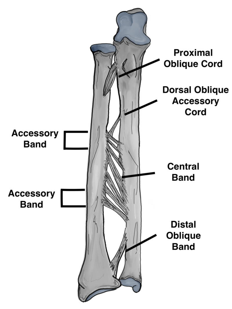
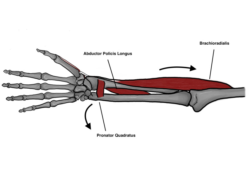
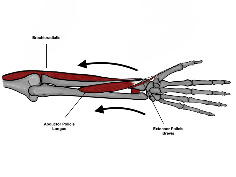
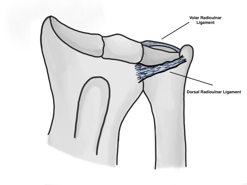
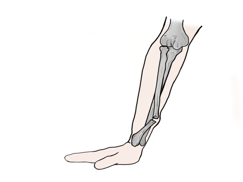
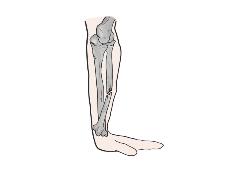
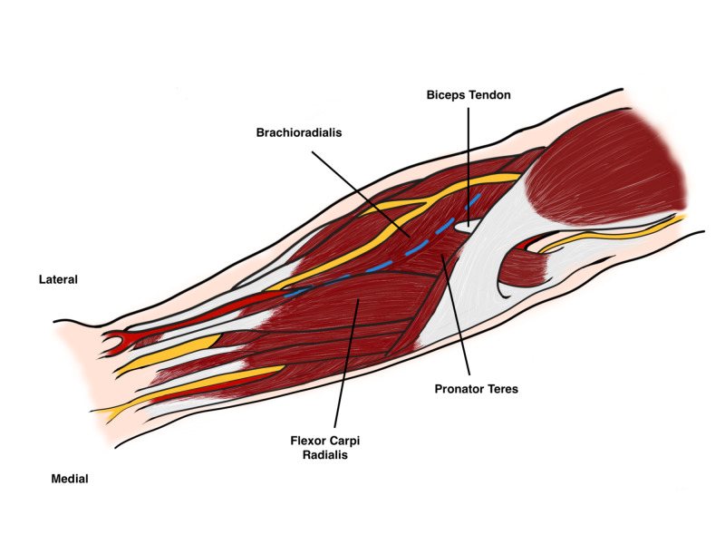
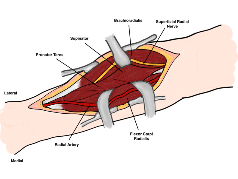
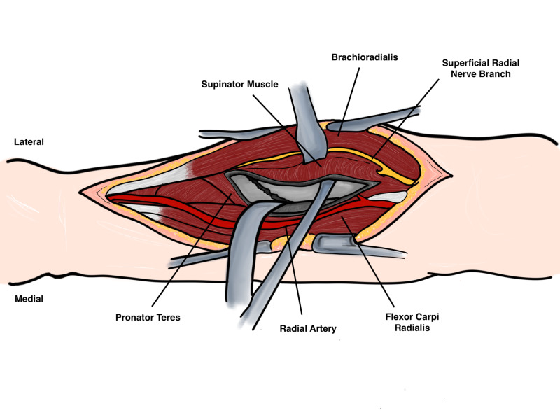
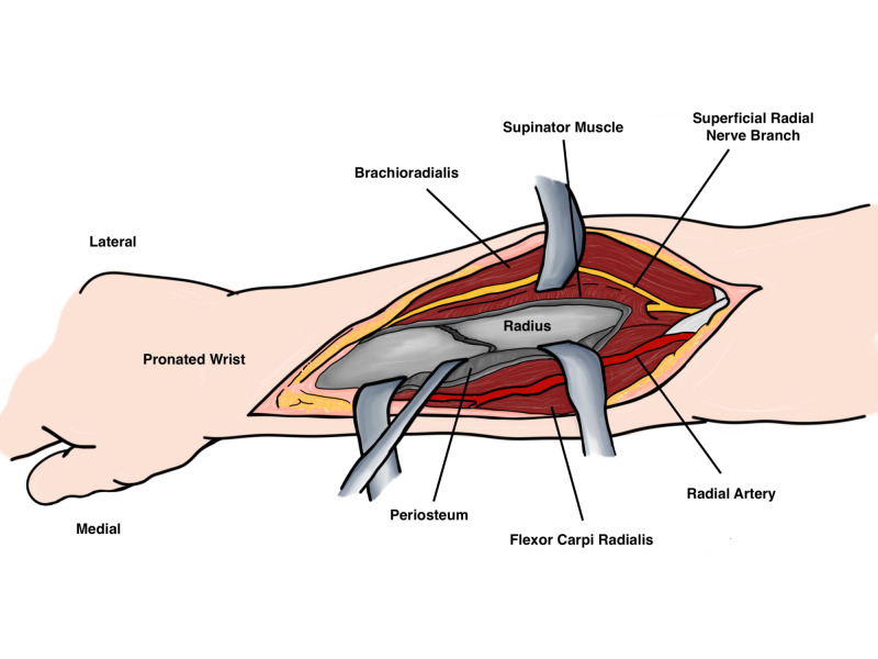
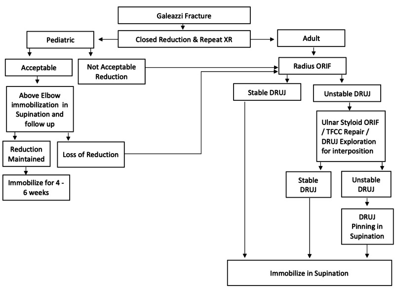
Galeazzi fracture dislocations are a fracture of the distal one third of the radius shaft with a concomitant dislocation of the distal radioulnar joint (DRUJ). These injuries usually occur by axial loading on an outstretched arm with pronation or supination of the wrist which determines the angulation of the fracture. Surgical treatment has been historically by the anterior (volar) approach to the forearm with plate fixation with or without pinning of the distal radioulnar joint. Failed or inadequate treatment may lead to complications including chronic pain, malunion or instability of the DRUJ that may warrant salvage procedures.

Keywords: distal radioulnar joint; fracture dislocation; galeazzi; radius fracture; tfcc.


Conflict of interest statement

The authors have declared that no competing interests exist.

Figures

Figure 1
Figure 1. Components of the interosseous membrane
Original illustration by Alswaji G.F.
Figure 2
Figure 2. Deforming forces acting on the distal fragment
Original illustration by Alswaji G.F.
Figure 3
Figure 3. Deforming forces acting on the distal fragment
Original illustration by Alswaji G.F.
Figure 4
Figure 4. Distal radioulnar ligaments
Original illustration by Alswaji G.F.
Figure 5
Figure 5. Apex volar galeazzi fracture
Original illustration by Alswaji G.F.
Figure 6
Figure 6. Apex dorsal galeazzi fracture
Original illustration by Alswaji G.F.
Figure 7
Figure 7. Diagnostic algorithm
Proposed by the author.
Figure 8
Figure 8. Anterior volar incision (Henry's Approach)
Original illustration by Alswaji G.F.
Figure 9
Figure 9. Superficial dissection
Original illustration by Alswaji G.F.
Figure 10
Figure 10. Deep dissection
Original illustration by Alswaji G.F.
Figure 11
Figure 11. Deep dissection
Original illustration by Alswaji G.F.
Figure 12
Figure 12. Treatment algorithm
Proposed by the author.

References

    1. A historical report on Riccardo Galeazzi and the management of Galeazzi fractures. Sebastin SJ, Chung KC. J Hand Surg Am. 2010;35:1870–1877. - PMC - PubMed
    1. Galeazzi lesions in children and adolescents: treatment and outcome. Eberl R, Singer G, Schalamon J, Petnehazy T, Hoellwarth ME. Clin Orthop Relat Res. 2008;466:1705–1709. - PMC - PubMed
    1. Galeazzi fractures and dislocations. Giannoulis FS, Sotereanos DG. Hand Clin. 2007;23:153–163. - PubMed
    1. Interosseous membrane and its effect on the distal radioulnar joint. Slutsky DJ. J Wrist Surg. 2016;5:171. - PMC - PubMed
    1. Galeazzi fracture. Atesok KI, Jupiter JB, Weiss A-P. J Am Acad Orthop Surg. 2011;19:623–633. - PubMed

LinkOut - more resources