Case Reports
. 2020 Jul;32(111):243-247.
doi: 10.22038/ijorl.2020.40635.2330.

Schwannoma of the Brachial Plexus: A Rare Case Report

Affiliations
Case Reports

Schwannoma of the Brachial Plexus: A Rare Case Report

Shruti Ranjan et al. Iran J Otorhinolaryngol. 2020 Jul.

Abstract

Introduction: Brachial plexus schwannomas are extremely rare tumours of the head and neck region accounting for less than 5 % overall. Due to its rarity and anatomic complexity of the brachial plexus, schwannomas in this region present a diagnostic and surgical challenge to the surgeon.

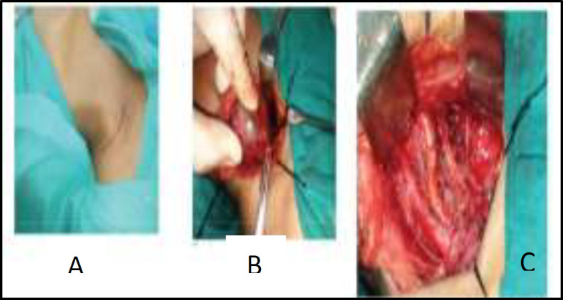
Case report: We present a case of a 56-year-old female who presented with a slow growing right sided neck swelling associated with pain and tingling in the distal end of the right forearm. According to FNAC, imaging studies results, a diagnosis of benign neurogenic tumour possibly schwannoma was made. After taking proper consent patient underwent surgical excision of the tumour. Postoperatively, patient developed numbness and tingling in right arm and stiffness at elbow joint, which is showing improvement after regular physiotherapy sessions.

Conclusion: Although brachial plexus schwannomas are extremely rare head and neck tumours they should be kept as a differential diagnosis in patients presenting with supraclavicular neck swellings. These are potentially curable lesions. As such, detailed history and examination together with imaging studies is important in establishing a preoperative diagnosis for proper management.

Keywords: Brachial plexus; Neurofibroma; Schwannomas.


Figures

Fig 1
Fig 1
Cect Neck: Well-defined heterogeneously enhancing solid cystic lesion in right supraclavicular region medially extending into and causing widening of right C6-C7 neural foramen
Fig 2
Fig 2
MRI Neck And Chest: Well defined solid cystic lesion in right supraclavicular region: (A) Isointense on T1WI; (B) Heterogenous enhancement seen on post contrast scan
Fig 3
Fig 3
(A) Horizontal incision over the swelling; (B) Trunks of the Brachial Plexus passing around the tumour; (C) Brachial Plexus after tumour removal
Fig 4
Fig 4
Characteristic cellular pattern of alternating regions containing compact spindle cells called Antoni type A areas and more loosely arranged, hypocellular zones called Antoni type B areas

References

    1. Ansari I, Ansari A, Graison AA, Patil AJ, Joshi H. Head and Neck Schwannomas: A Surgical Challenge-A Series of 5 Cases. Case Reports in Otolaryngology. 2018 Mar;:1–10. - PMC - PubMed
    1. Ryu KH, Moon JI, Baek HJ, Cho SB, Choi BH, An HJ et al. Brachial plexus schwannoma mimicking cervical lymphadenopathy: A case report with emphasis on imaging features. Medicine (Baltimore) 2018 Oct;97:42. - PMC - PubMed
    1. Rashid M, Salahuddin O, Yousaf S, Qazi UA, Yousaf K. Schwannoma of the brachial plexus; report of two cases involving the C7 root. J Brachial Plex Peripher Nerve Inj. 2013 Nov;8(1) - PMC - PubMed
    1. Kumar A, Akhtar S. Schwannoma of brachial plexus. Indian J Surg. 2011 Jan;73(1):80–1. - PMC - PubMed
    1. Vishwanathan N, Suma Devi B. Unusual cervical mass - A surgical dilemma and unfortunate sequelae. Indian J Otolaryngol Head Neck Surg. 2006 Jul;58(3):275–6. - PMC - PubMed

Publication types

LinkOut - more resources