Quantitative Assessment of Parenchymal Involvement Using 3D Lung Model in Adolescent With Covid-19 Interstitial Pneumonia
- PMID: 32850560
- PMCID: PMC7419575
- DOI: 10.3389/fped.2020.00453
Quantitative Assessment of Parenchymal Involvement Using 3D Lung Model in Adolescent With Covid-19 Interstitial Pneumonia
Abstract
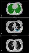
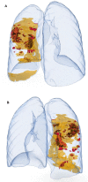
Background: Amount of parenchymal involvement in patients with interstitial pneumonia Covid-19 related, seems to be associated with a worse prognosis. Nowadays 3D reconstruction imaging is expanding its role in clinical medical practice. We aimed to use 3D lung reconstruction of a young lady affected by Sars-CoV2 infection and interstitial pneumonia, to better visualize, and quantitatively assess the parenchymal involvement. Methods: Volumetric Chest CT scan was performed in a 15 years old girl with interstitial lung pneumonia, Sars-CoV2 infection related. 3D modeling of the lungs, with differentiation of healthy and affected parenchymal area were obtained by using multiple software. Results: 3D reconstruction imaging allowed us to quantify the lung parenchyma involved, Self-explaining 3D images, useful for the understanding, and discussion of the clinical case were also obtained. Conclusions: Quantitative Assessment of Parenchymal Involvement Using 3D Lung Model in Covid-19 Infection is feasible and it provides information which could play a role in the management and risk stratification of these patients.
Keywords: 3D in Covid19; 3D lung reconstructions; 3D modeling in pneumonia; 3D parenchima reconstruction; 3D quantify in pneumonia; 3D rendering in pneumonia.
Copyright © 2020 Borro, Ciliberti, Santangelo, Magistrelli, Campana, Carducci, Caterina, Tomà and Secinaro.
Figures


References
-
- Sánchez-Sánchez Á, Girón-Vallejo Ó, Ruiz-Pruneda R, Fernandez-Ibieta M, García-Calderon D, Villamil V, et al. . Three-Dimensional printed model and virtual reconstruction: an extra tool for pediatric solid tumors surgery. Eur J Pediatr Surg Reports. (2018) 6:e70–6. 10.1055/s-0038-1672165 - DOI - PMC - PubMed
LinkOut - more resources
Full Text Sources
Miscellaneous

