

The Mechanics of Mitotic Cell Rounding
- PMID: 32850812
- PMCID: PMC7423972
- DOI: 10.3389/fcell.2020.00687
The Mechanics of Mitotic Cell Rounding
Abstract
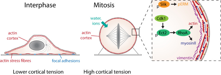
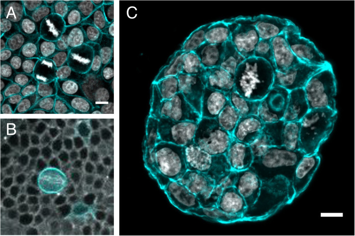
When animal cells enter mitosis, they round up to become spherical. This shape change is accompanied by changes in mechanical properties. Multiple studies using different measurement methods have revealed that cell surface tension, intracellular pressure and cortical stiffness increase upon entry into mitosis. These cell-scale, biophysical changes are driven by alterations in the composition and architecture of the contractile acto-myosin cortex together with osmotic swelling and enable a mitotic cell to exert force against the environment. When the ability of cells to round is limited, for example by physical confinement, cells suffer severe defects in spindle assembly and cell division. The requirement to push against the environment to create space for spindle formation is especially important for cells dividing in tissues. Here we summarize the evidence and the tools used to show that cells exert rounding forces in mitosis in vitro and in vivo, review the molecular basis for this force generation and discuss its function for ensuring successful cell division in single cells and for cells dividing in normal or diseased tissues.
Keywords: Ect2; actin cortex; cell mechanics; ezrin; mitosis; mitotic rounding; myosin; osmotic pressure.
Copyright © 2020 Taubenberger, Baum and Matthews.
Figures
Similar articles
-
Hydrostatic pressure and the actomyosin cortex drive mitotic cell rounding.Nature. 2011 Jan 13;469(7329):226-30. doi: 10.1038/nature09642. Epub 2011 Jan 2. Nature. 2011. PMID: 21196934
-
Genome-scale single-cell mechanical phenotyping reveals disease-related genes involved in mitotic rounding.Nat Commun. 2017 Nov 2;8(1):1266. doi: 10.1038/s41467-017-01147-6. Nat Commun. 2017. PMID: 29097687 Free PMC article.
-
F-Actin Interactome Reveals Vimentin as a Key Regulator of Actin Organization and Cell Mechanics in Mitosis.Dev Cell. 2020 Jan 27;52(2):210-222.e7. doi: 10.1016/j.devcel.2019.12.011. Epub 2020 Jan 9. Dev Cell. 2020. PMID: 31928973 Free PMC article.
-
Coupling changes in cell shape to chromosome segregation.Nat Rev Mol Cell Biol. 2016 Aug;17(8):511-21. doi: 10.1038/nrm.2016.75. Epub 2016 Jun 29. Nat Rev Mol Cell Biol. 2016. PMID: 27353479 Review.
-
Shaping up to divide: coordinating actin and microtubule cytoskeletal remodelling during mitosis.Semin Cell Dev Biol. 2014 Oct;34:109-15. doi: 10.1016/j.semcdb.2014.02.015. Epub 2014 Mar 4. Semin Cell Dev Biol. 2014. PMID: 24607328 Review.
Cited by
-
Actomyosin-Driven Division of a Synthetic Cell.ACS Synth Biol. 2022 Oct 21;11(10):3120-3133. doi: 10.1021/acssynbio.2c00287. Epub 2022 Sep 27. ACS Synth Biol. 2022. PMID: 36164967 Free PMC article. Review.
-
The role of RAS oncogenes in controlling epithelial mechanics.Trends Cell Biol. 2023 Jan;33(1):60-69. doi: 10.1016/j.tcb.2022.09.002. Epub 2022 Sep 27. Trends Cell Biol. 2023. PMID: 36175301 Free PMC article. Review.
-
Interplay between the plasma membrane and cell-cell adhesion maintains epithelial identity for correct polarised cell divisions.J Cell Sci. 2024 Mar 1;137(5):jcs261701. doi: 10.1242/jcs.261701. Epub 2023 Nov 28. J Cell Sci. 2024. PMID: 37888135 Free PMC article.
-
Cell Cycle Regulation by Integrin-Mediated Adhesion.Cells. 2022 Aug 14;11(16):2521. doi: 10.3390/cells11162521. Cells. 2022. PMID: 36010598 Free PMC article. Review.
-
Activity-dependent glassy cell mechanics Ⅰ: Mechanical properties measured with active microrheology.Biophys J. 2023 May 16;122(10):1781-1793. doi: 10.1016/j.bpj.2023.04.011. Epub 2023 Apr 11. Biophys J. 2023. PMID: 37050875 Free PMC article.
References
Publication types
Grants and funding
LinkOut - more resources
Full Text Sources