

New Insights Into Cranial Synchondrosis Development: A Mini Review
- PMID: 32850826
- PMCID: PMC7432265
- DOI: 10.3389/fcell.2020.00706
New Insights Into Cranial Synchondrosis Development: A Mini Review
Abstract
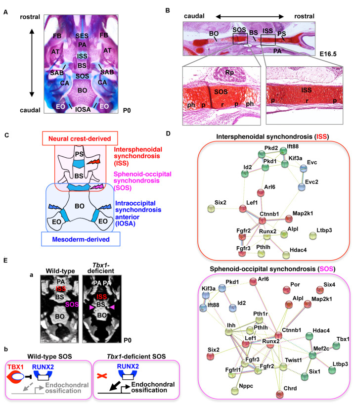
The synchondroses formed via endochondral ossification in the cranial base are an important growth center for the neurocranium. Abnormalities in the synchondroses affect cranial base elongation and the development of adjacent regions, including the craniofacial bones. In the central region of the cranial base, there are two synchondroses present-the intersphenoid synchondrosis and the spheno-occipital synchondrosis. These synchondroses consist of mirror image bipolar growth plates. The cross-talk of several signaling pathways, including the parathyroid hormone-like hormone (PTHLH)/parathyroid hormone-related protein (PTHrP), Indian hedgehog (Ihh), Wnt/β-catenin, and fibroblast growth factor (FGF) pathways, as well as regulation by cilium assembly and the transcription factors encoded by the RUNX2, SIX1, SIX2, SIX4, and TBX1 genes, play critical roles in synchondrosis development. Deletions or activation of these gene products in mice causes the abnormal ossification of cranial synchondrosis and skeletal elements. Gene disruption leads to both similar and markedly different abnormalities in the development of intersphenoid synchondrosis and spheno-occipital synchondrosis, as well as in the phenotypes of synchondroses and skeletal bones. This paper reviews the development of cranial synchondroses, along with its regulation by the signaling pathways and transcription factors, highlighting the differences between intersphenoid synchondrosis and spheno-occipital synchondrosis.
Keywords: RUNX2; cartilage; cranial base; intersphenoid synchondrosis; mesoderm; neural crest; spheno-occipital synchondrosis.
Copyright © 2020 Funato.
Figures
References
-
- Al Kaissi A., Ben Chehida F., Kenis V., Ganger R., Radler C., Hofstaetter J. G., et al. (2013). Broad spectrum of skeletal malformation complex in patients with cleidocranial dysplasia syndrome: radiographic and tomographic study. Clin. Med. Insights Arthritis Musculoskelet. Disord. 6 45–55. 10.4137/CMAMD.S11933 - DOI - PMC - PubMed
-
- Caparrós-Martín J. A., Valencia M., Reytor E., Pacheco M., Fernandez M., Perez-Aytes A., et al. (2013). The ciliary Evc/Evc2 complex interacts with smo and controls hedgehog pathway activity in chondrocytes by regulating Sufu/Gli3 dissociation and Gli3 trafficking in primary cilia. Hum. Mol. Genet. 22 124–139. 10.1093/hmg/dds409 - DOI - PubMed
Publication types
LinkOut - more resources
Full Text Sources
Research Materials