. 2020 Aug 11:7:185.
doi: 10.3389/fmolb.2020.00185. eCollection 2020.

Conjugative Coupling Proteins and the Role of Their Domains in Conjugation, Secondary Structure and in vivo Subcellular Location

Affiliations

Conjugative Coupling Proteins and the Role of Their Domains in Conjugation, Secondary Structure and in vivo Subcellular Location

Itxaso Álvarez-Rodríguez et al. Front Mol Biosci. .

Abstract

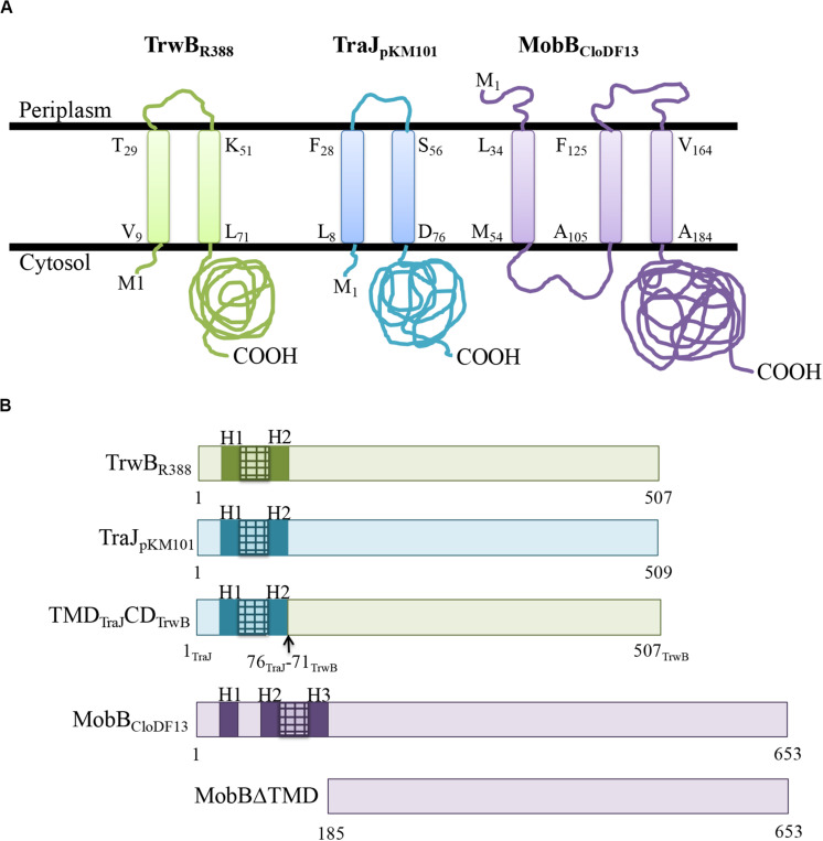
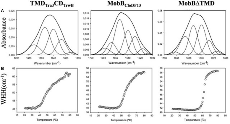
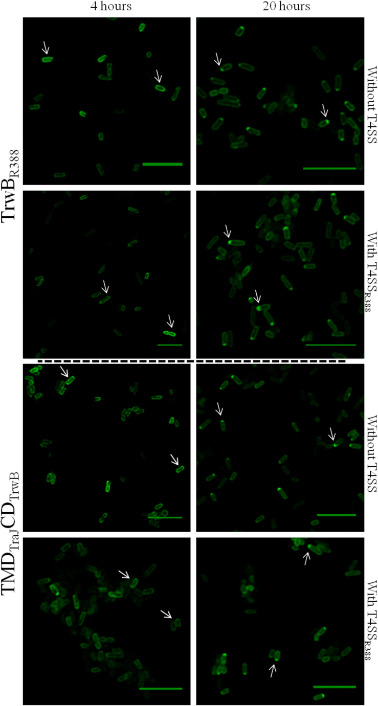
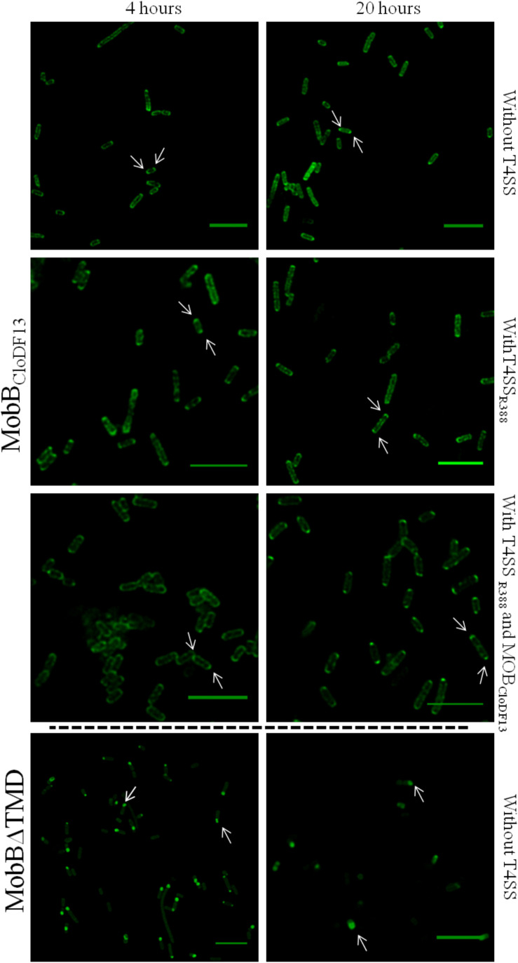
Type IV Coupling Proteins (T4CPs) are essential elements in many type IV secretion systems (T4SSs). The members of this family display sequence, length, and domain architecture heterogeneity, being the conserved Nucleotide-Binding Domain the motif that defines them. In addition, most T4CPs contain a Transmembrane Domain (TMD) in the amino end and an All-Alpha Domain facing the cytoplasm. Additionally, a few T4CPs present a variable domain at the carboxyl end. The structural paradigm of this family is TrwBR388, the T4CP of conjugative plasmid R388. This protein has been widely studied, in particular the role of the TMD on the different characteristics of TrwBR388. To gain knowledge about T4CPs and their TMD, in this work a chimeric protein containing the TMD of TraJpKM101 and the cytosolic domain of TrwBR388 has been constructed. Additionally, one of the few T4CPs of mobilizable plasmids, MobBCloDF13 of mobilizable plasmid CloDF13, together with its TMD-less mutant MobBΔTMD have been studied. Mating studies showed that the chimeric protein is functional in vivo and that it exerted negative dominance against the native proteins TrwBR388 and TraJpKM101. Also, it was observed that the TMD of MobBCloDF13 is essential for the mobilization of CloDF13 plasmid. Analysis of the secondary structure components showed that the presence of a heterologous TMD alters the structure of the cytosolic domain in the chimeric protein. On the contrary, the absence of the TMD in MobBCloDF13 does not affect the secondary structure of its cytosolic domain. Subcellular localization studies showed that T4CPs have a unipolar or bipolar location, which is enhanced by the presence of the remaining proteins of the conjugative system. Unlike what has been described for TrwBR388, the TMD is not an essential element for the polar location of MobBCloDF13. The main conclusion is that the characteristics described for the paradigmatic TrwBR388 T4CP should not be ascribed to the whole T4CP family. Specifically, it has been proven that the mobilizable plasmid-related MobBCloDF13 presents different characteristics regarding the role of its TMD. This work will contribute to better understand the T4CP family, a key element in bacterial conjugation, the main mechanism responsible for antibiotic resistance spread.

Keywords: antibiotic resistance spread; bacterial conjugation; coupling proteins; membrane proteins; type IV secretion systems.


Figures

FIGURE 1
FIGURE 1
(A) Predicted membrane topology of TrwBR388, TraJpKM101, and MobBCloDF13 proteins. Membrane topology of the different T4CPs was predicted using Topcons software. The black lines represent the inner bacterial membrane. M1, amino-terminus; COOH, carboxy-terminus. The first and last residues of each transmembrane helix are shown indicating their position in the sequence. Proteins from R388, pKM101, and CloDF13 plasmids are shown in green, blue, and purple, respectively. (B) Schematic representation of the different T4CPs and their variants used in the present study. Proteins from R388, pKM101, and CloDF13 plasmids are shown in green, blue and purple, respectively. The transmenbrane α-helices (H) and the small periplasmic loops connecting α-helices are indicated in dark boxes and stripped boxes, respectively.
FIGURE 2
FIGURE 2
(A) Amide I region of the infrared spectra of TMDTraJCDTrwB, MobBCloDF13, and MobBΔTMD. Proteins were purified, dialyzed against the corresponding buffer in D2O and analyzed by IR spectroscopy as explained in “Materials and Methods” section. Obtained spectra were curve-fitted to show the different secondary structure components as detailed in Table 5. (B) Thermal denaturation of TMDTraJCDTrwB, MobBCloDF13, and MobBΔTMD as seen by IR spectroscopy. The widths at half-height (WHH) of the amide I bands are plotted as a function of temperature (°C). Thermal denaturation is marked by an abrupt increase in bandwidth. Mid-point denaturation temperature (Tm) values corresponding to each protein are detailed in Table 5.
FIGURE 3
FIGURE 3
Subcellular location of TrwBR388GFP and TMDTraJCDTrwBGFP fusion-proteins by confocal fluorescence microscopy. Proteins were expressed in E. coli BL21C41 (DE3) strain by induction with 1 mM IPTG for 4 (left panels) and 20 h (right panels) at 25°C. Subcellular location of TrwBR388GFP and TMDTraJCDTrwBGFP was determined in E. coli strains containing plasmid pSU1456 that expresses all R388 conjugative proteins except TrwBR388, or without plasmid pSU1456. The images were acquired in a Leica TCS SP5 confocal fluorescence microscope, with a 60× oil immersion objective. Sample excitation was performed with 488 nm wavelength, while fluorescence emission was measured between 505 and 525 nm. The images were analyzed using Huygens and ImageJ softwares. Arrrowheads indicate the eGFP fluorescence through the periphery (1st column) and at a single cell pole (2nd column). Scale bar: 5 μm.
FIGURE 4
FIGURE 4
Subcellular location of MobBCloDF13GFP and MobBΔTMDGFP fusion-proteins by confocal fluorescence microscopy. MobBCloDF13GFP and MobBΔTMDGFP proteins were expressed in E. coli BL21C41 (DE3) and E. coli BL21 (DE3) strains, respectively. Subcellular location of these proteins was determined by induction with 1 mM IPTG after 4 (left panels) and 20 h (right panels) at 25°C. Additionally, subcelular location was analyzed in the presence of pSU1456 plasmid, which expresses all R388 conjugative proteins except TrwBR388, or without plasmid pSU1456. MobBCloDF13 was also expressed in the presence of both pSU1456 and pSU4833 that codes for the mobilization proteins of plasmid CloDF13 (MOBCloDF13), except for MobBCloDF13. The images were acquired in a Leica TCS SP5 confocal fluorescence microscope, with a 60× oil immersion objective. Sample excitation was performed with 488 nm wavelength, while fluorescence emission was measured between 505 and 525 nm. The images were analyzed using Huygens and ImageJ software. Arrrowheads indicate the eGFP fluorescence foci at both cell poles (MobBCloDF13) and at a single cell pole (MobBΔTMD). Scale bar: 5 μm.

References

    1. Agopian A., Quetin M., Castano S. (2016). Structure and interaction with lipid membrane models of Semliki Forest virus fusion peptide. Biochim. Biophys. Acta Biomembr. 1858 2671–2680. 10.1016/j.bbamem.2016.07.003 - DOI - PMC - PubMed
    1. Agúndez L., Zárate-Pérez F., Meier A. F., Bardelli M., Llosa M., Escalante C. R., et al. (2018). Exchange of functional domains between a bacterial conjugative relaxase and the integrase of the human adeno-associated virus. PLoS One 13:e0200841. 10.1371/journal.pone.0200841 - DOI - PMC - PubMed
    1. Alvarez-Martinez C. E., Christie P. J. (2009). Biological diversity of prokaryotic type IV secretion systems. Microbiol. Mol. Biol. Rev. 73 775–808. 10.1128/mmbr.00023-09 - DOI - PMC - PubMed
    1. Andraka N., Sánchez-Magraner L., García-Pacios M., Goñi F. M., Arrondo J. L. R. (2017). The conformation of human phospholipid scramblase 1, as studied by infrared spectroscopy. Effects of calcium and detergent. Biochim. Biophys. Acta Biomembr. 1859 1019–1028. 10.1016/j.bbamem.2017.02.015 - DOI - PubMed
    1. Arrondo J. L., Goñi F. M. (1999). Structure and dynamics of membrane proteins as studied by infrared spectroscopy. Prog. Biophys. Mol. Biol. 72 367–405. 10.1016/s0079-6107(99)00007-3 - DOI - PubMed