

doi: 10.1007/s00270-020-02621-3.
Epub 2020 Aug 26.
Bronchial Artery Embolization Performed in COVID-19 Patients: Tolerance and Outcomes
Affiliations
- PMID: 32851427
- PMCID: PMC7449632
- DOI: 10.1007/s00270-020-02621-3
Item in Clipboard
Bronchial Artery Embolization Performed in COVID-19 Patients: Tolerance and Outcomes
Cardiovasc Intervent Radiol.
2020 Dec.
No abstract available
Conflict of interest statement
The authors declare that they have no conflict of interest.
Figures
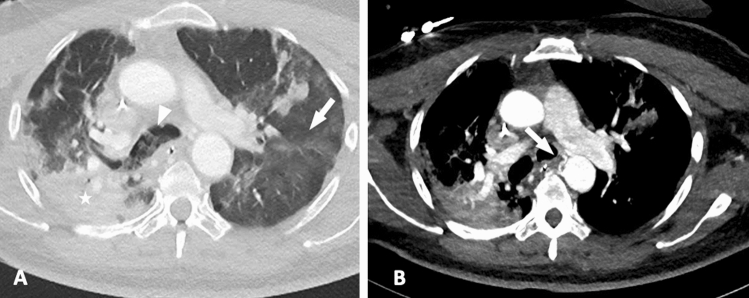
Contrast-enhanced chest computed tomography at the arterial phase in a 63-year-old patient with severe acute respiratory syndrome due to coronavirus 2019. A Chest CT demonstrates goggle-glass opacities with crazy-paving (white arrow), posterior condensation in the right upper lobe (white star) and a filling of the proximal right bronchus with hemorrhagic material (white arrowhead). B Chest CT demonstrates dilated bronchial arteries related to bronchial hypervascularization
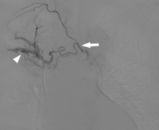
Digital subtractive angiography of right intercostobronchial artery. Selective catheterization demonstrates dilated bronchial artery with antegrade filling of the right upper lobe pulmonary artery, consistent with a bronchial–pulmonary arterial shunt
Similar articles
-
Clinical impact of multidetector row computed tomography before bronchial artery embolization in patients with hemoptysis: a prospective study.Can Assoc Radiol J. 2013 Feb;64(1):61-73. doi: 10.1016/j.carj.2011.08.002. Epub 2012 May 9. Can Assoc Radiol J. 2013. PMID: 22575595
-
Pediatric Racemose Hemangioma Treated with Angiographic Embolization.Am J Respir Crit Care Med. 2020 May 1;201(9):e70-e71. doi: 10.1164/rccm.201907-1449IM. Am J Respir Crit Care Med. 2020. PMID: 31930927 No abstract available.
-
[Imaging of actinomycosis: CT scan, bronchial embolization and pathology].Rev Mal Respir. 2024 Jun;41(6):446-450. doi: 10.1016/j.rmr.2024.05.002. Epub 2024 May 24. Rev Mal Respir. 2024. PMID: 38796385 French.
-
CT and CT angiography in massive haemoptysis with emphasis on pre-embolization assessment.Clin Radiol. 2011 Sep;66(9):869-75. doi: 10.1016/j.crad.2011.03.001. Epub 2011 Jun 11. Clin Radiol. 2011. PMID: 21658690 Review.
-
Radiological Evaluation and Endovascular Treatment of Hemoptysis.Curr Probl Diagn Radiol. 2016 May-Jun;45(3):215-24. doi: 10.1067/j.cpradiol.2015.07.007. Epub 2015 Jul 21. Curr Probl Diagn Radiol. 2016. PMID: 26293972 Review.
Cited by
-
CT evaluation of systemic artery to pulmonary artery fistula: an underdiagnosed disease in patients with hemoptysis.J Thorac Dis. 2023 Nov 30;15(11):5952-5960. doi: 10.21037/jtd-23-861. Epub 2023 Nov 2. J Thorac Dis. 2023. PMID: 38090324 Free PMC article.
-
Massive hemoptysis two months after an otherwise mild SARS-CoV-2 disease (COVID-19) treated with bronchial artery embolization - A case report.Radiol Case Rep. 2022 Mar;17(3):918-921. doi: 10.1016/j.radcr.2021.12.048. Epub 2022 Jan 15. Radiol Case Rep. 2022. PMID: 35069961 Free PMC article.
-
Obstructive Bronchial Fibrin Cast Formation in COVID-19 Severe Respiratory Failure.Am J Respir Crit Care Med. 2023 Feb 1;207(3):349-350. doi: 10.1164/rccm.202201-0225IM. Am J Respir Crit Care Med. 2023. PMID: 36174140 Free PMC article. No abstract available.
-
Retroperitoneal hemorrhage due to ruptured artery induced by median arcuate ligament syndrome in patients with COVID-19: A case series.Acute Med Surg. 2024 Nov 20;11(1):e70015. doi: 10.1002/ams2.70015. eCollection 2024 Jan-Dec. Acute Med Surg. 2024. PMID: 39575224 Free PMC article.
-
Automated Detection of Broncho-Arterial Pairs Using CT Scans Employing Different Approaches to Classify Lung Diseases.Biomedicines. 2023 Jan 5;11(1):133. doi: 10.3390/biomedicines11010133. Biomedicines. 2023. PMID: 36672641 Free PMC article.
References
Publication types
MeSH terms
LinkOut - more resources
Full Text Sources
Medical
