Comparative Study
. 2021 Feb;57(2):248-256.
doi: 10.1002/uog.22186.

Single-cell RNA expression profiling of SARS-CoV-2-related ACE2 and TMPRSS2 in human trophectoderm and placenta

Affiliations
Comparative Study

Single-cell RNA expression profiling of SARS-CoV-2-related ACE2 and TMPRSS2 in human trophectoderm and placenta

D Cui et al. Ultrasound Obstet Gynecol. 2021 Feb.

Abstract

Objectives: To examine the characteristics and distribution of possible severe acute respiratory syndrome coronavirus 2 (SARS-CoV-2) target cells in the human trophectoderm (TE) and placenta.

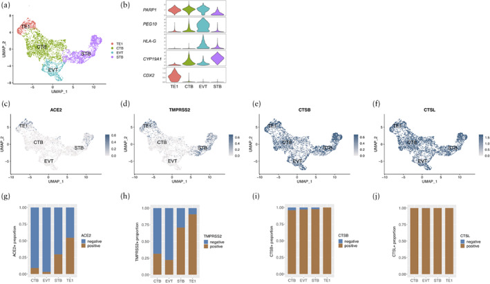
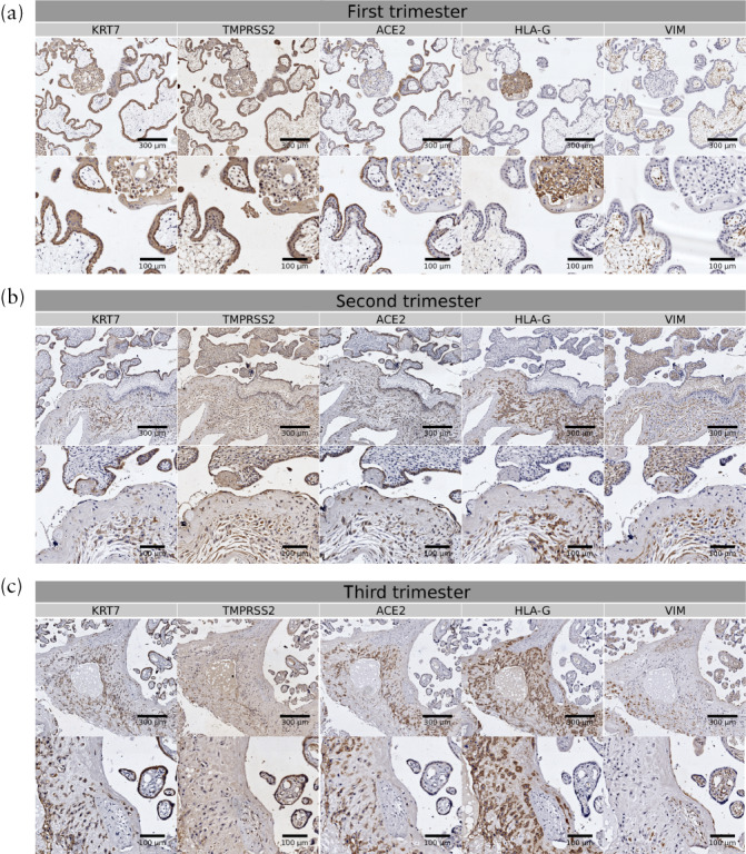
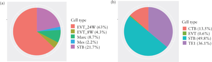
Methods: Bioinformatics analysis was performed based on published single-cell transcriptomic datasets of early TE and first- and second-trimester human placentae. We conducted the transcriptomic analysis of 4198 early TE cells, 1260 first-trimester placental cells and 189 extravillous trophoblast cells (EVTs) from 24-week placentae (EVT_24W) using the SMART-Seq2 method. In addition, to confirm the bioinformatic results, we performed immunohistochemical staining of three first-trimester, three second-trimester and three third-trimester placentae from nine women recruited prospectively to this study. We evaluated the expression of the SARS-CoV-2-related molecules angiotensin-converting enzyme 2 (ACE2) and transmembrane protease serine 2 (TMPRSS2).

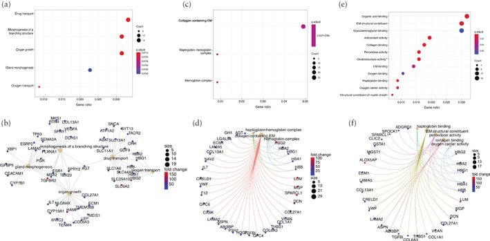
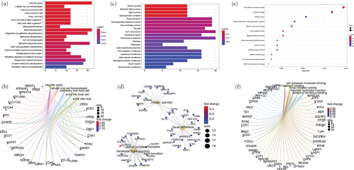
Results: Via bioinformatic analysis, we identified the existence of ACE2 and TMPRSS2 expression in human TE as well as in first- and second-trimester placentae. In the human TE, 54.4% of TE1 cells, 9.0% of cytotrophoblasts (CTBs), 3.2% of EVTs and 29.5% of syncytiotrophoblasts (STBs) were ACE2-positive. In addition, 90.7% of TE1 cells, 31.5% of CTBs, 22.1% of EVTs and 70.8% of STBs were TMPRSS2-positive. In placental cells, 20.4% of CTBs, 44.1% of STBs, 3.4% of EVTs from 8-week placentae (EVT_8W) and 63% of EVT_24W were ACE2-positive, while 1.6% of CTBs, 26.5% of STBs, 1.9% of EVT_8W and 20.1% of EVT_24W were TMPRSS2-positive. Pathway analysis revealed that EVT_24W cells that were positive for both ACE2 and TMPRSS2 (ACE2 + TMPRSS2-positive) were associated with morphogenesis of branching structure, extracellular matrix interaction, oxygen binding and antioxidant activity. The ACE2 + TMPRSS2-positive TE1 cells were correlated with an increased capacity for viral invasion, epithelial-cell proliferation and cell adhesion. Expression of ACE2 and TMPRSS2 was observed on immunohistochemical staining in first-, second- and third-trimester placentae.

Conclusions: ACE2- and TMPRSS2-positive cells are present in the human TE and placenta in all three trimesters of pregnancy, which indicates the possibility that SARS-CoV-2 could spread via the placenta and cause intrauterine fetal infection. © 2020 International Society of Ultrasound in Obstetrics and Gynecology.

Keywords: ACE2; COVID-19; SARS-CoV-2; TMPRSS2; placenta; trophectoderm.


Figures

Figure 1
Figure 1
(a,b) Uniform manifold approximation and projection (UMAP) distributions showing cell clusters (a) and violin plots displaying expression levels of representative markers in each cell type (b) of single‐cell RNA sequencing data from embryonic trophectoderm (TE) cells obtained at 6–14 days after fertilization. Cells were classified into four clusters: trophectoderm 1 (TE1), syncytiotrophoblasts (STB), extravillous trophoblasts (EVT) and cytotrophoblasts (CTB) (c–f). Expression in early TE cells of SARS‐CoV‐2‐related molecules: angiotensin‐converting enzyme 2 (ACE2) (c), transmembrane protease serine 2 (TMPRSS2) (d), cathepsin B (CTSB) (e) and cathepsin L (CTSL) (f). (g–j) Proportion of cells that were positive for ACE2 (g), TMPRSS2 (h), CTSB (i) and CTSL (j) according to cell type.
Figure 2
Figure 2
(a,b) t‐distributed stochastic neighbor embedding (t‐SNE) distributions showing cell clusters (a) and violin plots displaying expression levels of representative markers in each cell type (b) of single‐cell RNA sequencing data from 8‐week and 24‐week placentae. Cells were classified into seven clusters: blood, cytotrophoblasts (CTB), extravillous trophoblasts from 8‐week (EVT_8W) and 24‐week (EVT_24W) placentae, macrophages (Marc), mesenchymal cells (Mes) and syncytiotrophoblasts (STB) (c–f). Expression in first‐ and second‐trimester placentae of SARS‐CoV‐2‐related molecules angiotensin‐converting enzyme 2 (ACE2) (c), transmembrane protease serine 2 (TMPRSS2) (d), cathepsin B (CTSB) (e) and cathepsin L (CTSL) (f). (g–j) Proportion of cells that were positive for ACE2 (g), TMPRSS2 (h), CTSB (i) and CTSL (j) according to cell type.
Figure 3
Figure 3
Immunohistochemical staining of angiotensin‐converting enzyme 2 (ACE2) and transmembrane protease serine 2 (TMPRSS2) in first‐ (a), second‐ (b) and third‐ (c) trimester human placentae. Staining of keratin 7 (KRT7; marker of trophoblasts), human leukocyte antigen G (HLA‐G; marker of extravillous trophoblasts) and vimentin (VIM; marker of stromal cells) was also conducted to show different cell types. Scale bars in upper rows of figure parts represent 300 µm and those in lower rows represent 100 µm.
Figure 4
Figure 4
Distribution of cells that were positive for both angiotensin‐converting enzyme 2 (ACE2) and transmembrane protease serine 2 (TMPRSS2) in 8‐week (8W) and 24‐week (24W) human placentae (a) and in early trophectoderm (b), across cell types. CTB, cytotrophoblasts; EVT, extravillous trophoblasts; Marc, macrophages; Mes, mesenchymal cells; STB, syncytiotrophoblasts; TE1, trophectoderm 1.
Figure 5
Figure 5
Gene ontology (GO) analysis showing enriched terms in extravillous trophoblasts from 24‐week human placentae (EVT_24W) that were positive for both angiotensin‐converting enzyme 2 and transmembrane protease serine 2 in comparison to rest of EVT_24W cells that were not, including GO biological process analysis (a,b), GO cellular component analysis (c,d) and GO molecular function analysis (e,f). Colors in dot plots (a,c,e) are coded according to significance of enrichment scores and sizes are on basis of counts of overlapping genes. In network plots (b,d,f), size of each dot represents count of overlapping genes and color of each gene dot represents significance of enrichment scores. *Acting on peroxide as acceptor. EM, extracellular matrix.
Figure 6
Figure 6
Gene ontology (GO) analysis of upregulated genes in trophectoderm 1 (TE1) cells from early embryo that were positive for both angiotensin‐converting enzyme 2 and transmembrane protease serine 2 in comparison to rest of TE1 cells that were not, including GO biological process analysis (a,b), GO cellular component analysis (c,d) and GO molecular function analysis (e,f). Colors in bar plots (a,c) are coded according to significance of enrichment scores and heights are on basis of overlapping genes. Colors in dot plot (e) are coded according to significance of enrichment scores and sizes are on basis of counts of overlapping genes. In network plots (b,d,f), size of each dot represents count of overlapping genes and color of each gene dot represents significance of enrichment scores. *Involved in symbiotic interaction. TRA, transcription repressor activity; TTA, transmembrane transporter activity.

References

    1. Wang C, Chen D, Yang H. Updates on COVID‐19 Infection. Matern Med 2020; 2: 65–67. - PMC - PubMed
    1. Maltepe E, Fisher SJ. Placenta: the forgotten organ. Annu Rev Cell Dev Biol 2015; 31: 523–552. - PubMed
    1. Pereira L. Congenital Viral Infection: Traversing the Uterine‐Placental Interface. Annu Rev Virol 2018; 5: 273–299. - PubMed
    1. Arora N, Sadovsky Y, Dermody TS, Coyne CB. Microbial Vertical Transmission during Human Pregnancy. Cell Host Microbe 2017; 21: 561–567. - PMC - PubMed
    1. Lan J, Ge J, Yu J, Shan S, Zhou H, Fan S, Zhang Q, Shi X, Wang Q, Zhang L, Wang X. Structure of the SARS‐CoV‐2 spike receptor‐binding domain bound to the ACE2 receptor. Nature 2020; 581: 215–220. - PubMed

Publication types

MeSH terms