

Cerebral blood flow in dystonia due to pantothenate kinase-associated neurodegeneration
- PMID: 32851917
- PMCID: PMC7788684
- DOI: 10.1177/1971400920943967
Cerebral blood flow in dystonia due to pantothenate kinase-associated neurodegeneration
Abstract
Background and purpose: The aim of this study was to look for deviations of cerebral perfusion in patients suffering from pantothenate kinase-associated neurodegeneration, where the globus pallidus is affected by severe accumulation of iron.
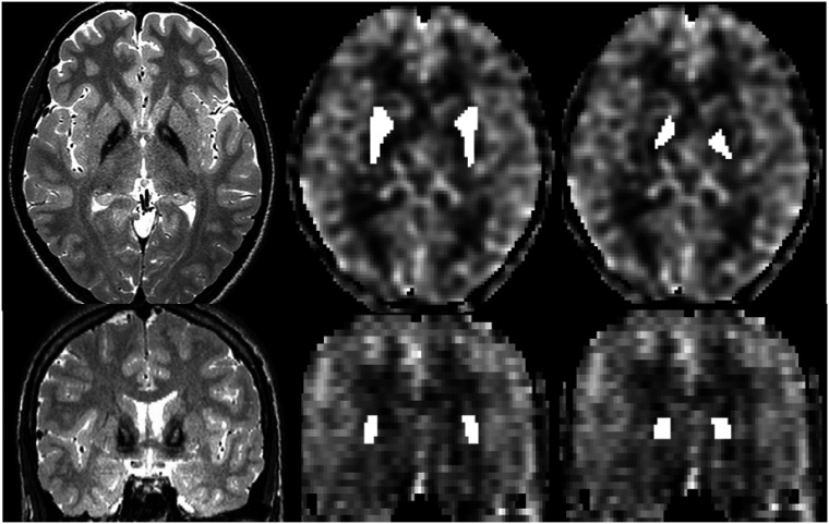
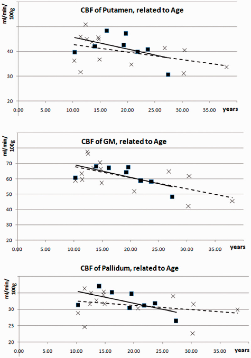
Material and methods: Under resting conditions, cerebral blood flow was measured by the magnetic resonance imaging technique of arterial spin labelling in cortical areas and basal ganglia in eight pantothenate kinase-associated neurodegeneration patients and 14 healthy age-matched control subjects and correlated to T2* time of these areas and - in patients - to clinical parameters.
Results: Despite highly significant differences of T2* time of the globus pallidus (20 vs 39 ms, p < 0.001), perfusion values of this nucleus were nearly identical in both groups (32 ± 3.3 vs 31 ± 4.0 ml/min/100 g) as well as in total brain gray matter (both 62 ± 6.7 resp. ±10.3 ml/min/100 g), putamen (41 ± 5.4 vs 40 ± 6.1 ml/min/100 g), in selected cortical regions, and the cerebellum. Correlations between perfusion and T2* time to clinical data did not reach significance (p > 0.05).
Conclusion: The absence of any obvious deviations of perfusion in the group of patients during a resting condition does not support the view that (non-functional) vascular pathology is a major pathogenic factor in pantothenate kinase-associated neurodegeneration in the younger age group. The findings underline the value of the arterial spin technique to measure cerebral blood flow in areas of disturbed susceptibility.
Keywords: Pantothenate kinase-associated neurodegeneration; cerebral blood flow; globus pallidus; tiger’s eye.
Figures
References
-
- Hayflick SJ, Westaway SK, Levinson B, et al. Genetic, clinical, and radiographic delineation of Hallervorden-Spatz syndrome. N Engl J Med 2003; 348: 33–40. - PubMed
-
- Vilchez-Abreu C, Roa-Sanchez P, Fermin-Delgado R, et al. El signo del “Ojo del Tigre” en resonancia magnética: Cambios relacionados con la edad. Anal Radiol Mexico 2013; 3: 189–196.
MeSH terms
LinkOut - more resources
Full Text Sources
