. 2018 Jan-Dec:17:1536012118808644.
doi: 10.1177/1536012118808644.

Current Practice and Emerging Molecular Imaging Technologies in Oral Cancer Screening

Affiliations

Current Practice and Emerging Molecular Imaging Technologies in Oral Cancer Screening

Arianna Strome et al. Mol Imaging. 2018 Jan-Dec.

Abstract

Oral cancer is one of the most common cancers globally. Survival rates for patients are directly correlated with stage of diagnosis; despite this knowledge, 60% of individuals are presenting with late-stage disease. Currently, the initial evaluation of a questionable lesion is performed by a conventional visual examination with white light. If a lesion is deemed suspicious, a biopsy is taken for diagnosis. However, not all lesions present suspicious under visual white light examination, and there is limited specificity in differentiating between benign and malignant transformations. Several vital dyes, light-based detection systems, and cytology evaluation methods have been formulated to aid in the visualization process, but their lack of specific biomarkers resulted in high false-positive rates and thus limits their reliability as screening and guidance tools. In this review, we will analyze the current methodologies and demonstrate the need for specific intraoral imaging agents to aid in screening and diagnosis to identify patients earlier. Several novel molecular imaging agents will be presented as, by result of their molecular targeting, they aim to have high specificity for tumor pathways and can support in identifying dysplastic/cancerous lesions and guiding visualization of biopsy sites. Imaging agents that are easy to use, inexpensive, noninvasive, and specific can be utilized to increase the number of patients who are screened and monitored in a variety of different environments, with the ultimate goal of increasing early detection.

Keywords: conventional oral examination (COE); molecular markers; molecularly targeted approaches; oral squamous cell carcinoma (OSCC); specificity; vital dyes.


Conflict of interest statement

Declaration of Conflicting Interests: The author(s) declared the following potential conflicts of interest with respect to the research, authorship, and/or publication of this article. T.R. is a cofounder of Summit Biomedical Imaging, LLC. S.K., S.P., and T.R. are shareholders of Summit Biomedical Imaging, LLC. Dr. Milind Rajadhyaksha is a former employee of and owns equity in Caliber I.D. (formerly, Lucid Inc.), the company that manufactures and sells a reflectance confocal microscope (VivaScope). The VivaScope, for reflectance confocal microscopy (RCM) imaging, is the commercial version of an original laboratory prototype that was developed by Dr. Rajadhyaksha when he was at Massachusetts General Hospital, Harvard Medical School.

Figures

Figure 1.
Figure 1.
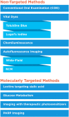
Overview of imaging methods discussed in the article.
Figure 2.
Figure 2.
Morphological appearance of different pathologies on the tongue. Malignant (a) and benign (b) oral lesions can be difficult to distinguish with white light alone.
Figure 3.
Figure 3.
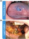
Vital dyes as diagnostic adjuncts. (a) Toluidine blue staining the oral cavity to identify leukoplakia lesion. (b) Lugol’s iodine staining oral mucosa, labels correspond to the following, A: invasive squamous cell carcinoma, B: dysplastic tissue, C: normal mucosa, D: normal orthokeratinized mucosa. Absence of staining in A indicates the malignant nature of the tissue. Adapted from McMahon et al and Awan et al.
Figure 4.
Figure 4.
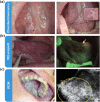
Light-based detection systems as diagnostic adjuncts. (a) Mild dysplasia on the ventral tongue, left: white light, right: chemiluminescence. (b) Severe dysplasia on the ventral tongue, left: white light, right: wide-field autofluorescence imaging with VELscope with arrow pointing to the dysplasia. (c) Left: oral squamous cell carcinoma on the hard palate, right: reflectance confocal microscopy demonstrating disorganized cells. (a) and (b) are macroscopic techniques; (c) is a microscopic technique. Adapted from Epstein et al, Shin et al, and Contaldo et al.
Figure 5.
Figure 5.
Fluorescence-based diagnostic adjuncts. (a) Wheat germ agglutinin-FITC staining in the oral cavity of a patient with OSCC, left: white light, right: WGA-FITC, A: squamous cell carcinoma with a high level of differentiation between normal tissue and malignancy by increased WGA-FITC uptake, C: moderate dysplasia can display an increased or decreased WGA-FITC uptake. The example shows good differentiation from normal tissue by reduction of WGA-FITC uptake. (b) Widefield fluorescence images of 3 oral specimens obtained pre and post labeling with 2-NBDG. Neoplastic samples showed lower fluorescence pre-labeling than normal samples and a dramatic increase of fluorescence after labeling. (c) Protoporphyrin IX-based OSCC imaging in combination with autofluorescence. A: moderately differentiated squamous cell carcinoma under white light, B: wide-field autofluorescence demonstrating loss of fluorescence in the malignant area, C: 5-ALA induced PpIX fluorescence with strong red fluorescence signal from the tumor area, D: overlayed autofluorescence and PpIX images resulting in a “street light” green/red contrast of tumor to the surrounding tissue. (d) Schematic of the imaging procedure for oral cancer detection using a PARPi-FL based, orally applied solution in an ongoing phase I/II clinical study (NCT03085147) (left). Proof of principle of feasibility of tumor detection after topical application of PARPi-FL (right). Adapted from Baeten et al, Betz et al, Nitin et al, and Kossatz et al.5-ALA, 5-aminolevulinic acid; 2-NBDG, 2-(N-(7-nitrobenz-2-oxa-1,3-diazol-4-yl)amino)-2-deoxyglucose; OSCC, oral squamous cell carcinoma; PpIX, Protoporphyrin IX; WGA, wheat germ agglutinin.

Similar articles

Cited by

References

    1. Bagan J, Sarrion G, Jimenez Y. Oral cancer: clinical features. Oral Oncol. 2010;46(6):414–417. - PubMed
    1. Le Campion A, Ribeiro CMB, Luiz RR, et al. Low survival rates of oral and oropharyngeal squamous cell carcinoma. Int J Dent. 2017;2017:5815493. - PMC - PubMed
    1. Warnakulasuriya S. Global epidemiology of oral and oropharyngeal cancer. Oral Oncol. 2009;45(4-5):309–316. - PubMed
    1. Ferlay J, Shin HR, Bray F, Forman D, Mathers C, Parkin DM. Estimates of worldwide burden of cancer in 2008: GLOBOCAN 2008. Int J Cancer. 2010;127(12):2893–2917. - PubMed
    1. Shield KD, Ferlay J, Jemal A, et al. The global incidence of lip, oral cavity, and pharyngeal cancers by subsite in 2012. CA Cancer J Clin. 2017;67(1):51–64. - PubMed

LinkOut - more resources