. 2021 Jul 27;117(9):2083-2091.
doi: 10.1093/cvr/cvaa253.

Mechanism of ventricular premature beats elicited by left stellate ganglion stimulation during acute ischaemia of the anterior left ventricle

Affiliations

Mechanism of ventricular premature beats elicited by left stellate ganglion stimulation during acute ischaemia of the anterior left ventricle

Bastiaan J D Boukens et al. Cardiovasc Res. .

Abstract

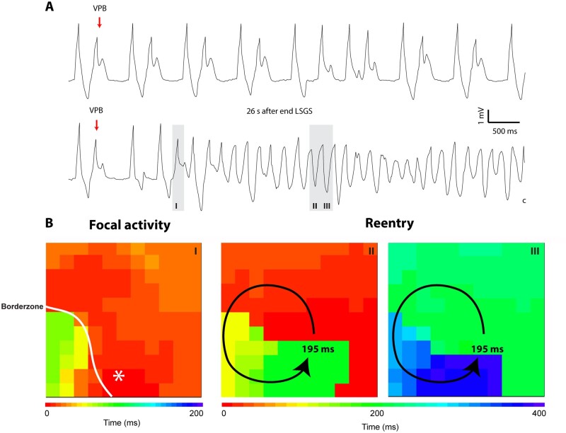
Aims: Enhanced sympathetic activity during acute ischaemia is arrhythmogenic, but the underlying mechanism is unknown. During ischaemia, a diastolic current flows from the ischaemic to the non-ischaemic myocardium. This 'injury' current can cause ventricular premature beats (VPBs) originating in the non-ischaemic myocardium, especially during a deeply negative T wave in the ischaemic zone. We reasoned that shortening of repolarization in myocardium adjacent to ischaemic myocardium increases the 'injury' current and causes earlier deeply negative T waves in the ischaemic zone, and re-excitation of the normal myocardium. We tested this hypothesis by activation and repolarization mapping during stimulation of the left stellate ganglion (LSG) during left anterior descending coronary artery (LAD) occlusion.

Methods and results: In nine pigs, five subsequent episodes of acute ischaemia, separated by 20 min of reperfusion, were produced by occlusion of the LAD and 121 epicardial local unipolar electrograms were recorded. During the third occlusion, left stellate ganglion stimulation (LSGS) was initiated after 3 min for a 30-s period, causing a shortening of repolarization in the normal myocardium by about 100 ms. This resulted in more negative T waves in the ischaemic zone and more VPBs than during the second, control, occlusion. Following the decentralization of the LSG (including removal of the right stellate ganglion and bilateral cervical vagotomy), fewer VPBs occurred during ischaemia without LSGS. During LSGS, the number of VPBs was similar to that recorded before decentralization.

Conclusion: LSGS, by virtue of shortening of repolarization in the non-ischaemic myocardium by about 100 ms, causes deeply negative T waves in the ischaemic tissue and VPBs originating from the normal tissue adjacent to the ischaemic border.

Keywords: Arrhythmias; Autonomic nervous system; Injury current; Ischaemia; Repolarization.


Figures

None
Graphical abstract
Figure 1
Figure 1
Schematic representation of the left ventricle with the multiple electrode covering both the ischaemic tissue (blue) and the non-ischaemic myocardium. The left stellate ganglion innervates primarily the area outside the ischaemic zone (red). LSGS, left stellate ganglion stimulation.
Figure 2
Figure 2
Diagram showing hypothetical transmembrane action potentials in normal (red) and ischaemic tissue (blue) (left), and local extracellular electrograms from the ischaemic myocardium (right). In the middle, diagrams showing intra- and extracellular current flow at the moments indicated by the dotted lines drawn through the action potentials at the left and the local electrograms at the right., Before LSGS (upper panels), this current causes a current source in the extracellular space in the ischaemic tissue leading to a positive T wave. During LSGS (lower panels), the action potential in the non-ischaemic zone shortens, and the current now causes a current sink and a negative T wave in the ischaemic tissue.
Figure 3
Figure 3
(A) ST elevation in the ischaemic zone and ST depression in the non-ischaemic tissue. (B) Repolarization maps before (top left panel) and after 30 s of LSGS (top right panel) without ischaemia. The bottom left panel shows the repolarization map after 3 min of ischaemia. The bottom right panels show the repolarization maps after 30 s of LSGS during continued ischaemia. Note shortening of repolarization time in the non-ischaemic zone in the order of 100 ms. The bar graphs show repolarization times in the anterior and lateral wall during LSGS without (C) and during ischaemia (D). Data are shown as mean ± SEM (n = 8). It was not possible to determine repolarization times in the anterior wall during ischaemia because of ST-segment elevation. (C) *P = 0.014 interaction effect LSGS*location (two-way ANOVA, LSGS = repeated factor, location = repeated factor) followed by a Bonferroni-corrected t-test (P = 0.0018). (D) *P = 0.0031 Bonferroni-corrected t-test. LSGS, left stellate ganglion stimulation.
Figure 4
Figure 4
(A) Superimposed local electrograms from normal and ischaemic tissue after 3 min and 3′30″ of ischaemia. Note the development of a negative T wave in the ischaemic zone when repolarization outside the ischaemic zone is markedly shortened by LSGS (B). (C) T-wave amplitude of local electrograms recorded from the ischaemic zone on after 3 min and 3′30″ of ischaemia, with (red) and without (blue) LSGS. Data are shown as mean ± SEM (n = 8). *P = 0.021 interaction effect LSGS*location (two-way ANOVA, LSGS = repeated factor, location = repeated factor) followed by a Bonferroni-corrected t-test (P = 0.006).
Figure 5
Figure 5
Local electrograms from the ischaemic zone after 3 min of ischaemia without (upper trace) and with LSGS (lower trace). Without LSGS, no VPB’s occur. With LSGS, negative T waves develop, which are followed by VPB’s. Note alternans of negative T waves in (A) (bottom). VPB, ventricular premature beat.
Figure 6
Figure 6
Local electrograms and activation map of a single VPB (A and B). The VPB originated in the non-ischaemic myocardium close to the ischaemic border (a) and followed a deeply negative T wave in the ischaemic zone (b). (C) The average number (±SEM) of VPBs in seven pigs, after various times of ischaemia with LSGS (red bars) and without LSGS (blue). Data are shown both with an intact denervation and following decentralization of the LSG, removal of the RSG, and bilateral vagotomy. *P = 0.019 main effect of LSGS on the occurrence of VPBs (three-way Analysis of Variance, decentralization = repeated factor, LSGS = repeated factor, time = repeated factor). The effect of LSGS on VPBs was significant before and after correcting (log transformation) for possible skewedness of the data. (D) Incidence of ventricular tachycardia (VT) or ventricular fibrillation (VF) in eight pigs. These arrhythmias only occurred after LSGS (P = 0.015, chi-square 5.9). LSGS, left stellate ganglion stimulation; RSG, right stellate ganglion ;VPB, ventricular premature beat.
Figure 7
Figure 7
Local electrograms during the control occlusion (A upper trace) showing bigeminy, and after the third occlusion with LSGS when ventricular fibrillation occurred (A lower trace). In B, activation maps of the first VPB that initiated ventricular fibrillation, showing a focal activity, and beats 11 and 12 of VF showing reentry. VPB, ventricular premature beat.

Similar articles

Cited by

References

    1. Verrier RL, Lown B.. Influence of neural activity on ventricular electrical stability during acute myocardial ischemia and infarction. In Sandoe E., Julian D.G., Bell J.W. (eds) Management of Ventricular Tachycardia—Role of Mexilitine. Amsterdam: Excerpta Medica, 1978. pp. 133–150.
    1. Corr PB, Yamada KA, Witkowski FX.. Mechanisms controlling cardiac autonomic function and their relation to arrhythmogenesis. In Fozzard F.A., Haber E., Jennings R.B., Katz A.M., Morgan H.E. (eds) The Heart and Cardiovascular System. New York: Raven Press, 1986. pp. 1343–1404.
    1. Ajijola OA, Vaseghi M, Mahajan A, Shivkumar K.. Bilateral cardiac sympathetic denervation: why, who and when. Expert Rev Cardiovasc Ther 2012;10:947–949. - PMC - PubMed
    1. Vaseghi M, Barwad P, Malavassi Corrales FJ, Tandri H, Mathuria N, Shah R, Sorg JM, Gima J, Mandal K, Sàenz Morales LC, Lokhandwala Y, Shivkumar K.. Sympathetic denervation for refractory ventricular arrhythmias. J Am Coll Cardiol 2017;69:3070–3080. - PMC - PubMed
    1. Schwartz PJ. Cardiac sympathetic denervation to prevent life-threatening arrhythmias. Nat Rev Cardiol 2014;11:346–353. - PubMed

Publication types